. 2018 Sep;24(9):1317-1323.
doi: 10.1038/s41591-018-0106-7. Epub 2018 Jul 16.

Fetal gene therapy for neurodegenerative disease of infants

Affiliations

Fetal gene therapy for neurodegenerative disease of infants

Giulia Massaro et al. Nat Med. 2018 Sep.

Abstract

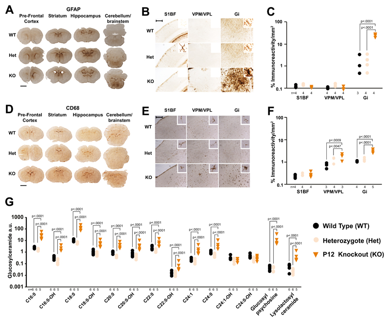
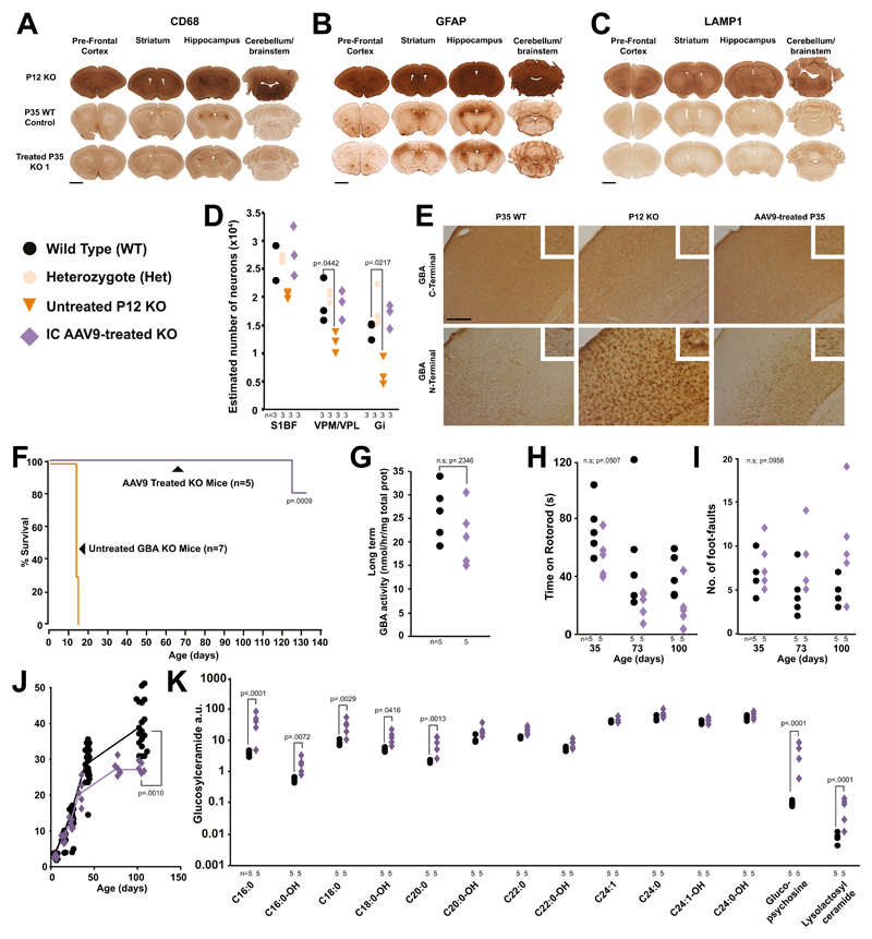
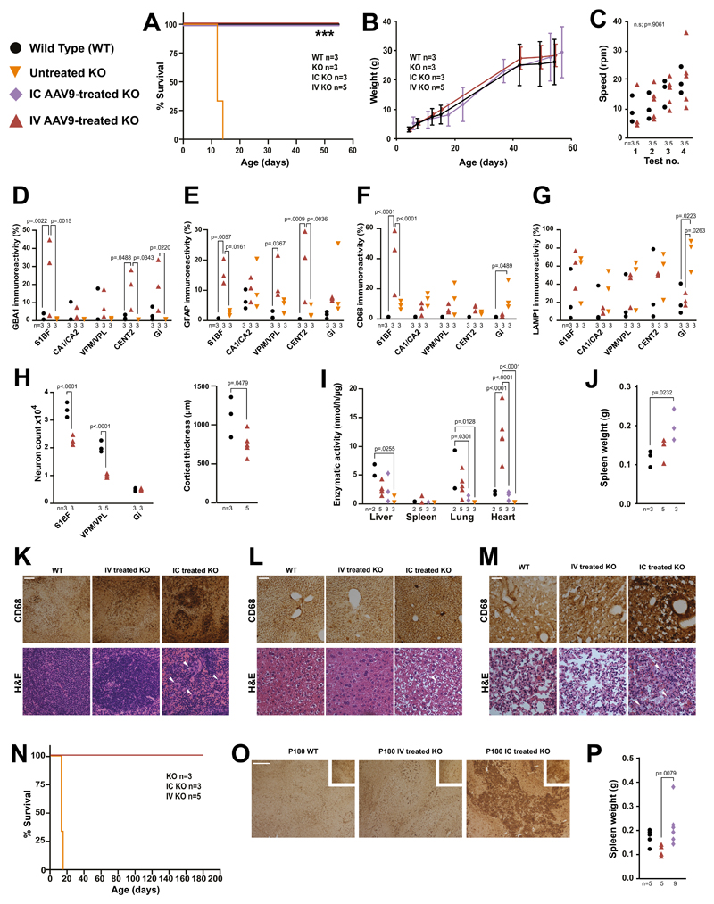
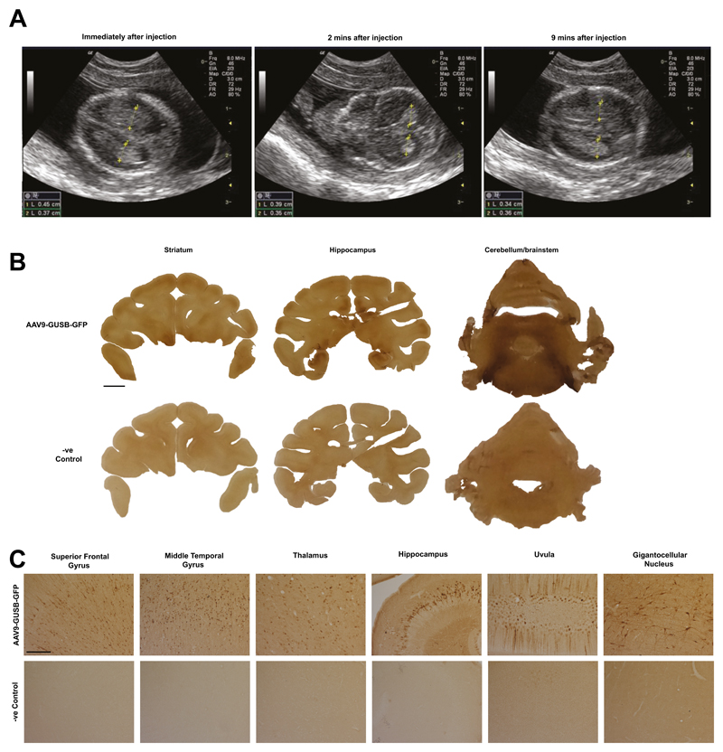
For inherited genetic diseases, fetal gene therapy offers the potential of prophylaxis against early, irreversible and lethal pathological change. To explore this, we studied neuronopathic Gaucher disease (nGD), caused by mutations in GBA. In adult patients, the milder form presents with hepatomegaly, splenomegaly and occasional lung and bone disease; this is managed, symptomatically, by enzyme replacement therapy. The acute childhood lethal form of nGD is untreatable since enzyme cannot cross the blood-brain barrier. Patients with nGD exhibit signs consistent with hindbrain neurodegeneration, including neck hyperextension, strabismus and, often, fatal apnea1. We selected a mouse model of nGD carrying a loxP-flanked neomycin disruption of Gba plus Cre recombinase regulated by the keratinocyte-specific K14 promoter. Exclusive skin expression of Gba prevents fatal neonatal dehydration. Instead, mice develop fatal neurodegeneration within 15 days2. Using this model, fetal intracranial injection of adeno-associated virus (AAV) vector reconstituted neuronal glucocerebrosidase expression. Mice lived for up to at least 18 weeks, were fertile and fully mobile. Neurodegeneration was abolished and neuroinflammation ameliorated. Neonatal intervention also rescued mice but less effectively. As the next step to clinical translation, we also demonstrated the feasibility of ultrasound-guided global AAV gene transfer to fetal macaque brains.


Conflict of interest statement

Conflicts of interest

Seng H. Cheng is an employee at Sanofi, a biopharmaceutical company

Figures

Fig. 1
Fig. 1. Brain disease at birth in nGD mice.
(A) GFAP immunostaining in newborn knockouts, heterozygous and WT brains. Scale bar = 1mm (B) Higher magnification of (A) in the somatosensory barrel field cortex (S1BF), ventral posteromedial/posterolateral nuclei (VPM/VPL) and gigantocellular nucleus (Gi). Scale bar = 0.25mm. (C) Quantification of (B) (2-way ANOVA, Tukey’s multiple comparison). (D) CD68 immunohistochemistry in newborn knockout, heterozygous and WT brains. Scale bar = 1mm (E) Higher magnification of (D). (F) Quantification of (E) (2-way ANOVA). (G) Mass spectrometry analysis of glucosylceramide isoforms, glucopsychosine and lyso-lactosylceramide in knockouts, heterozygote and WT brains (2-way ANOVA on log-transformed data; Bonferroni’s multiple comparison). Numbers of mice are stated under each group. n.s. = not significant
Fig. 2
Fig. 2. Fetal gene therapy of nGD mice.
(A) CD68, (B) GFAP and (C) LAMP1 brain immunostaining of untreated post-gestational day 12 (P12) knockouts and P35 WT and treated mice from different litters (scale bar = 1mm). (D) Stereological estimates of neuron number in the brains of treated knockout, heterozygote, WT and untreated day 12 knockouts (2-way ANOVA, Bonferroni’s multiple comparison, all versus WT). (Representative of 3 KO replicates shown in Fig. S6) (E) GCase C-terminal antibody labelling neurons in WTs, knockouts and treated knockout brains. N-terminal antibody staining in WT and treated knockouts (S1BF images, scale bar = 0.25mm). Representative of 3 mice replicates per experimental cohort. (F) Survival studies of treated knockouts compared to untreated knockouts (Mantel-Cox test, n=5 and 7 mice, respectively). (G) GCase enzyme activity in treated and WT brains (Students one-tailed t-test). (H) Rotarod and (I) foot-fault tests on treated knockout and WT mice (2-way ANOVA, Bonferroni’s multiple comparison). (J) Weights of treated and WT mice over time (Students one-tailed t-test on weight at 100 days, WT n=19, KO n=5). (K) Mass spectrometry profiles in the brains of treated knockout and WT mice (2-way ANOVA on log-transformed data, Bonferroni’s multiple comparison). Numbers of mice are stated under each group. n.s. = not significant
Fig. 3
Fig. 3. Intracerebroventricular and intravenous gene therapy in neonatal nGD mice.
(A) Kaplan-Meier survival plot of untreated knockouts, IC gene therapy treated knockouts, IV gene therapy treated knockouts and WTs (Logrank, Mantel-Cox test). (B) Weights of mice from (A). (C) Rotarod assessment of mice from (A) (repeated measures ANOVA). Immunohistochemistry for GBA1 (D), GFAP (E), CD68 (F) and LAMP1 (G) in brains of 55 day old treated knockout and WTs and P12 untreated knockouts in the S1BF, hippocampal cornu ammonus 1 (CA1), the cerebellar central lobule II (CENT2) and Gi (2-way ANOVA, Tukey’s multiple comparisons). (H) Neuron counts in the Gi, S1BF and VPM/VPL in treated knockouts and WTs and cortical thickness measurements (2-way ANOVA, Tukey’s multiple comparisons). (I) GCase activity in organs of treated knockouts, WTs, and untreated P12 knockouts (2-way ANOVA, Tukey’s multiple comparisons). (J) Spleen weights from mice receiving gene therapy and WTs (1-way ANOVA, Tukey’s multiple comparisons). Haemotoxylin and eosin staining and CD68 immunohistochemistry of (K) spleens, (L) livers and (M) lungs from intravenously and intracerebroventricularly gene therapy treated and WT mice (white arrows highlight Gaucher cells, scale bar = 0.1mm). (N) Kaplan-Meier survival plot of long-term gene therapy treated and untreated knockouts (Mantel-Cox test). (O) CD68 immunohistochemistry on spleens from (N) and WTs (scale bar = 0.25mm). (P) Spleen weights from (O) (1-way ANOVA, Tukey’s multiple comparisons). Numbers of mice are stated under each group. n.s. = not significant
Fig. 4
Fig. 4. In utero gene delivery to the macaque brain via ultrasound-guided intracerebroventricular injection of vector.
(A) Ultrasound images following intracerebroventricular administration over time. (B) Green fluorescent protein immunohistochemistry on brain sections from administered macaques and control. Scale bar = 5mm. (C) Higher magnification examination of (B) Scale bar = 0.25mm. Representative of 2 administered macaques replicates

Comment in

References

    1. Gupta N, Oppenheim IM, Kauvar EF, Tayebi N, Sidransky E. Type 2 Gaucher disease: phenotypic variation and genotypic heterogeneity. Blood Cells Mol Dis. 2011;46:75–84. - PMC - PubMed
    1. Enquist IB, et al. Murine models of acute neuronopathic Gaucher disease. Proc Natl Acad Sci U S A. 2007;104:17483–17488. - PMC - PubMed
    1. Farfel-Becker T, et al. Spatial and temporal correlation between neuron loss and neuroinflammation in a mouse model of neuronopathic Gaucher disease. Hum Mol Genet. 2011 - PubMed
    1. Farfel-Becker T, et al. Neuronal accumulation of glucosylceramide in a mouse model of neuronopathic Gaucher disease leads to neurodegeneration. Hum Mol Genet. 2013 - PMC - PubMed
    1. Cabrera-Salazar MA, et al. Systemic delivery of a glucosylceramide synthase inhibitor reduces CNS substrates and increases lifespan in a mouse model of type 2 Gaucher disease. PLoS ONE. 2012;7:e43310. - PMC - PubMed

Publication types