. 2018 Jul 5:10:1901-1906.
doi: 10.2147/CMAR.S161871. eCollection 2018.

Safety and efficacy of denosumab in the treatment of pulmonary metastatic giant cell tumor of bone

Affiliations

Safety and efficacy of denosumab in the treatment of pulmonary metastatic giant cell tumor of bone

Yi Luo et al. Cancer Manag Res. .

Abstract

Background: Giant cell tumor (GCT) of bone is an intermittent and locally aggressive tumor with increasing pulmonary metastatic potential. In this study, we evaluated the interim clinical outcome of denosumab in patients with pulmonary metastatic GCT.

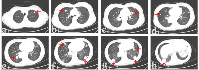
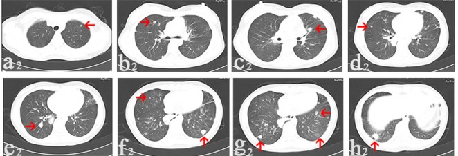
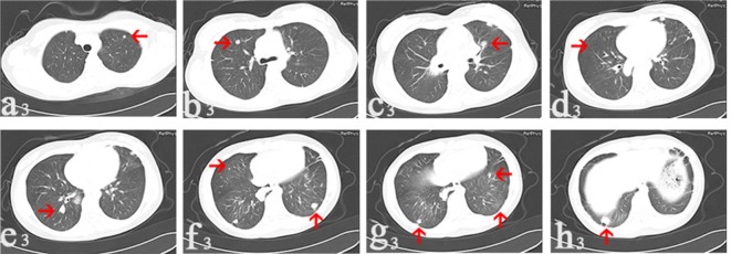
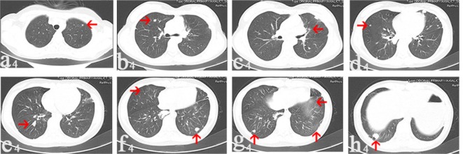
Materials and methods: We retrospectively reviewed seven patients with pulmonary metastatic GCT who received denosumab treatment after local tumor surgery during January 2014 and July 2016. Denosumab treatment for all patients lasted for at least 12 months. Serial chest computerized tomography scan was used to monitor the drug response and RECIST 1.1 standard was used to evaluate the therapeutic efficacy.

Results: All patients experienced chest pain relief in the first month of treatment. Three patients showed partial response. Four patients got stable disease after denosumab treatment. Adverse events included one patient with hypocalcemia and two patients with fever. No treatment-related deaths were reported. No patient with metastatic disease progression was found during an average of 28.6 months follow-up period.

Conclusion: We presented a promising interim clinical outcome using denosumab to treat patients with pulmonary metastatic GCT. Denosumab might be considered as the first-line treatment for patients with inoperable metastatic pulmonary GCT. However, Phase II clinical study with larger number of patients and longer follow-up period is needed to detect the further efficacy and safety of this drug for lung metastatic GCT.

Keywords: RANKL; denosumab; giant cell tumor of bone; pulmonary metastasis; recurrence.


Conflict of interest statement

Disclosure The authors report no conflicts of interest in this work.

Figures

Figure 1
Figure 1
In February 2014, CT scans of the third patient before denosumab treatment showed multi-lung metastatic nodules (arrows). Notes: The nodule was located on the left lung and grew to 1 cm in diameter (a1); the nodule was 1.2 cm in diameter (b1); the nodule was 1.2 cm in diameter (c1); the nodule was 0.6 cm in diameter (d1); the nodule was 2.2 cm in diameter (e1); the nodules were 1.2 and 2.2 cm in diameter (f1); two nodules were 2.2 cm and the other one was 1.2 cm in diameter (g1); the nodule was 2.4 cm in diameter (h1). Abbreviation: CT, computerized tomography.
Figure 2
Figure 2
In August 2014, CT images collected after denosumab treatment for 6 months showed multi-lung metastatic nodules (arrows) reduced in size and number. Notes: The nodule reduced to 0.4 cm in diameter (a2); the nodule reduced to 0.6 cm in diameter (b2); the nodule was with lower density than before and reduced to 1.0 cm in diameter (c2); the nodule reduced to 0.4 cm in diameter (d2); the nodule reduced to 1.2 cm in diameter (e2); the nodule on the right side reduced to 1.2 cm in diameter and the one on the left side disappeared (f2); one nodule disappeared and two nodules reduced to 1.0 and 0.8 cm in diameter (g2); the nodule reduced to 1.4 cm in diameter (h2). Abbreviation: CT, computerized tomography.
Figure 3
Figure 3
In March 2015, CT images collected after denosumab treatment for 13 months. Notes: The nodule remained stable in size (a3); the nodule remained stable with size 0.6 cm in diameter (b3); the diameter of the nodule decreased to 0.8 cm (c3); one nodule disappeared on the right side (d3); there was no change in the size of the nodule (e3); the one on the left side reduced to 1.0 cm in diameter (f3); the two nodules reduced to 0.8 and 0.6 cm in diameter, respectively (g3); the nodule reduced to 1.2 cm in diameter (h3). Arrows point to the nodules. Abbreviation: CT, computerized tomography.
Figure 4
Figure 4
In July 2016, there was no new measurable nodule (arrows) on CT images collected after denosumab treatment for 29 months. Notes: The nodule on the left side disappeared (g4). Number and size of these metastatic nodules maintain stability on all these eight images (a4, b4, c4, d4, e4, f4, h4), when compared with images in March 2015. Abbreviation: CT, computerized tomography.

References

    1. Beebe-Dimmer JL, Cetin K, Fryzek JP, Schuetze SM, Schwartz K. The epidemiology of malignant giant cell tumors of bone: an analysis of data from the Surveillance, Epidemiology and End Results Program (1975–2004) Rare Tumors. 2009;1(2):e52. - PMC - PubMed
    1. Mendenhall WM, Zlotecki RA, Scarborough MT, Gibbs CP, Mendenhall NP. Giant cell tumor of bone. Am J Clin Oncol. 2006;29(1):96–99. - PubMed
    1. Tandra VS, Kotha KM, Satyanarayana MG, Vadlamani KV, Yerravalli V. Synchronous multicentric giant cell tumour of distal radius and sacrum with pulmonary metastases. Case Rep Oncol Med. 2015;2015:354158. - PMC - PubMed
    1. Niu X, Zhang Q, Hao L, et al. Giant cell tumor of the extremity: retrospective analysis of 621 Chinese patients from one institution. J Bone Joint Surg Am. 2012;94(5):461–467. - PubMed
    1. Bertoni F, Present D, Sudanese A, Baldini N, Bacchini P, Campanacci M. Giant-cell tumor of bone with pulmonary metastases. Six case reports and a review of the literature. Clin Orthop Relat Res. 1988;237:275–285. - PubMed

LinkOut - more resources