. 2018 Jul 2:10:184.
doi: 10.3389/fnagi.2018.00184. eCollection 2018.

Predicting Age From Brain EEG Signals-A Machine Learning Approach

Affiliations

Predicting Age From Brain EEG Signals-A Machine Learning Approach

Obada Al Zoubi et al. Front Aging Neurosci. .

Abstract

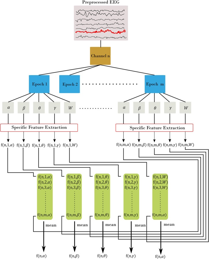
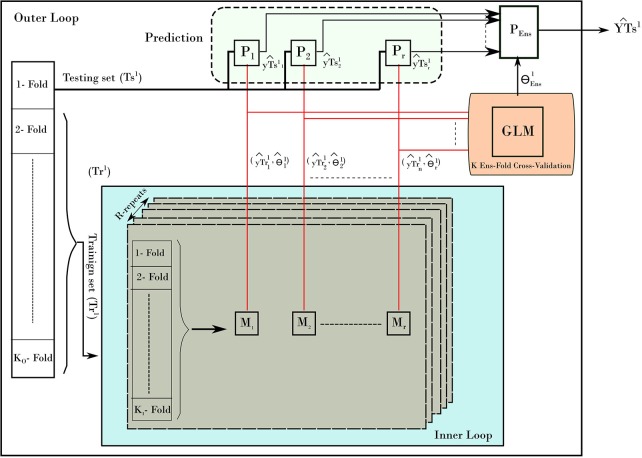
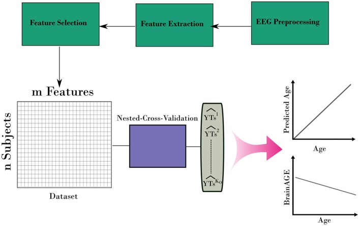
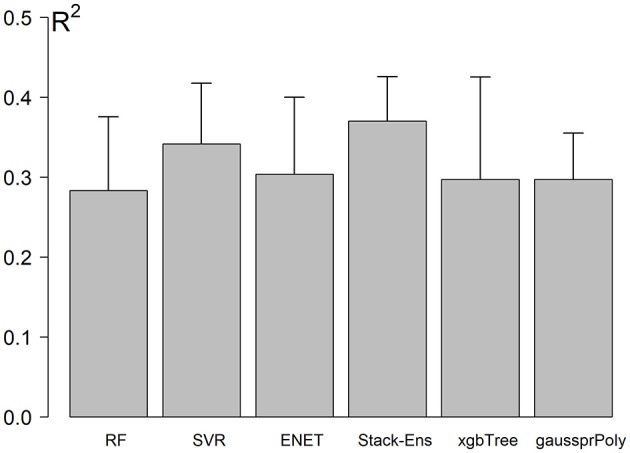
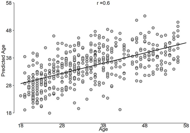
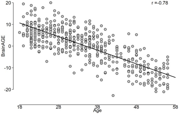
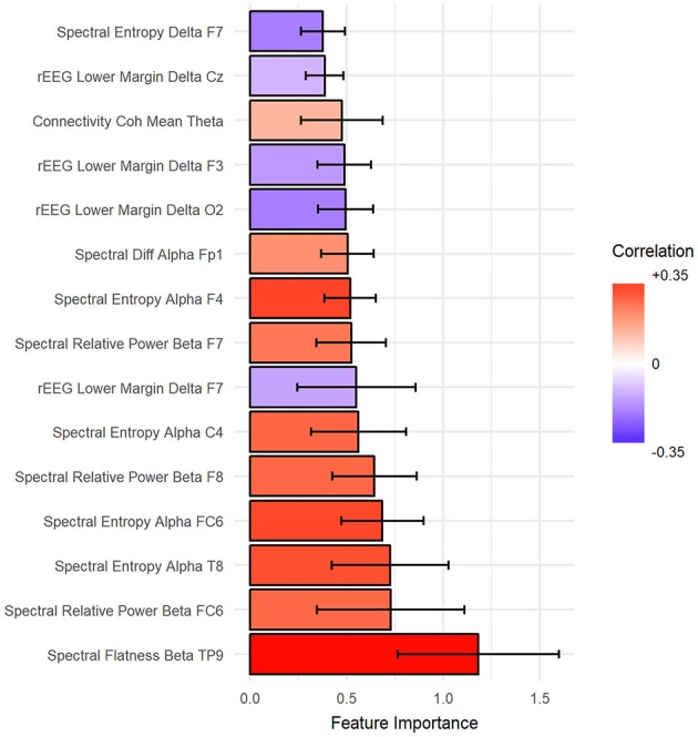
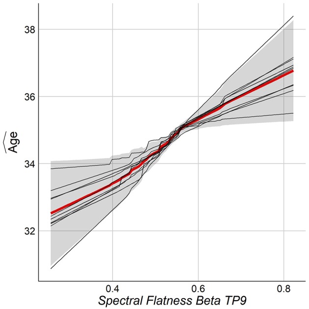
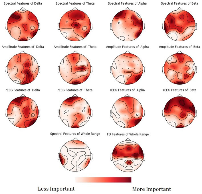
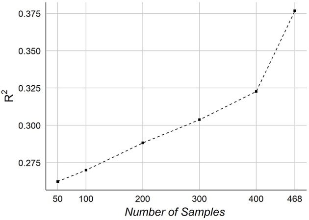
Objective: The brain age gap estimate (BrainAGE) is the difference between the estimated age and the individual chronological age. BrainAGE was studied primarily using MRI techniques. EEG signals in combination with machine learning (ML) approaches were not commonly used for the human age prediction, and BrainAGE. We investigated whether age-related changes are affecting brain EEG signals, and whether we can predict the chronological age and obtain BrainAGE estimates using a rigorous ML framework with a novel and extensive EEG features extraction. Methods: EEG data were obtained from 468 healthy, mood/anxiety, eating and substance use disorder participants (297 females) from the Tulsa-1000, a naturalistic longitudinal study based on Research Domain Criteria framework. Five sets of preprocessed EEG features across channels and frequency bands were used with different ML methods to predict age. Using a nested-cross-validation (NCV) approach and stack-ensemble learning from EEG features, the predicted age was estimated. The important features and their spatial distributions were deduced. Results: The stack-ensemble age prediction model achieved R2 = 0.37 (0.06), Mean Absolute Error (MAE) = 6.87(0.69) and RMSE = 8.46(0.59) in years. The age and predicted age correlation was r = 0.6. The feature importance revealed that age predictors are spread out across different feature types. The NCV approach produced a reliable age estimation, with features consistent behavior across different folds. Conclusion: Our rigorous ML framework and extensive EEG signal features allow a reliable estimation of chronological age, and BrainAGE. This general framework can be extended to test EEG association with and to predict/study other physiological relevant responses.

Keywords: BrainAGE; EEG; aging; feature extraction; human brain; machine learning.


Figures

Figure 1
Figure 1
Feature extraction procedure. Each channel is divided into m epoch. From there, we filtered each epoch into α, β, θ, γ, and W frequency bands. Then, for each filtered epoch, we applied the desired feature. This resulted in m feature value from all epochs, which are then averaged to estimate the channel-level feature. In the figure, we represent each feature using three indices: f(channel, epoch, band) with channel = [1.N], epoch = [1.m], and band = [α, β, θ, γ, W]. The final out is a channel-level feature and represented with two indices f(channel, band).
Figure 2
Figure 2
The nested-cross-validation procedure for predicting age. The example here demonstrates the first fold of the outer loop. The procedure consists of an inner loop (blue color) and outer loop. The inner loop is used to find the best models to predict the age. The outer loop uses those models to predict the age on the testing set. The process is repeated for all folds of the outer loop, which results in building a prediction of age from all samples.
Figure 3
Figure 3
The complete framework for estimating the BrainAGE form EEG. The framework uses the nested-cross-validation method (Figure 2) to build estimation for the age. Then, those estimations are used to calculate the BrainAGE from the entire dataset.
Figure 4
Figure 4
Models performance using NCV. Error bar represents the standard deviation of performances across the outer loop of NCV.
Figure 5
Figure 5
Predicted age vs. age constructed from the outer loop of NCV.
Figure 6
Figure 6
The BrainAGE variable as a function of the chronological age.
Figure 7
Figure 7
The top 15 important features to predict age sorted from most important (bottom) to top. Ventricle axis shows the scoring values from stack-ensemble model predictor, while the color indicates the correlation values between that feature and age.
Figure 8
Figure 8
PDP for the top feature from NCV from Stack-Ensemble model.
Figure 9
Figure 9
Mean feature importance scores sorted by bands and channels for predicting Age. The darker the color, the more important is the feature.
Figure 10
Figure 10
The effect of the number of samples on the age prediction.

References

    1. Allen P. J., Josephs O., Turner R. (2000). A method for removing imaging artifact from continuous EEG recorded during functional MRI. Neuroimage 12, 230–239. 10.1006/nimg.2000.0599 - DOI - PubMed
    1. Al Zoubi O., Awad M., Kasabov N. K. (2018). Anytime multipurpose emotion recognition from EEG data using a liquid state machine based framework. Artif. Intell. Med. 86, 1–8. 10.1016/j.artmed.2018.01.001 - DOI - PubMed
    1. Ashburner J. (2007). A fast diffeomorphic image registration algorithm. Neuroimage 38, 95–113. 10.1016/j.neuroimage.2007.07.007 - DOI - PubMed
    1. Babiloni C., Binetti G., Cassarino A., Dal Forno G., Del Percio C., Ferreri F., et al. (2006). Sources of cortical rhythms in adults during physiological aging: a multicentric EEG study. Hum. Brain Mapp. 27, 162–172. 10.1002/hbm.20175 - DOI - PMC - PubMed
    1. Babiloni C., Triggiani A. I., Lizio R., Cordone S., Tattoli G., Bevilacqua V., et al. (2016). Classification of single normal and Alzheimer's disease individuals from cortical sources of resting state EEG rhythms. Front. Neurosci. 10:47. 10.3389/fnins.2016.00047 - DOI - PMC - PubMed