. 2018 Apr 26:2018:8487248.
doi: 10.1155/2018/8487248. eCollection 2018.

Evaluation of the Protective Effect of Olive Leaf Extract on Cisplatin-Induced Testicular Damage in Rats

Affiliations

Evaluation of the Protective Effect of Olive Leaf Extract on Cisplatin-Induced Testicular Damage in Rats

Rafa S Almeer et al. Oxid Med Cell Longev. .

Abstract

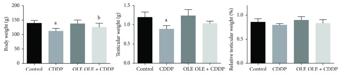
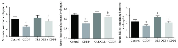
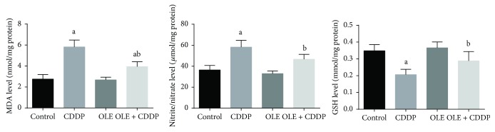
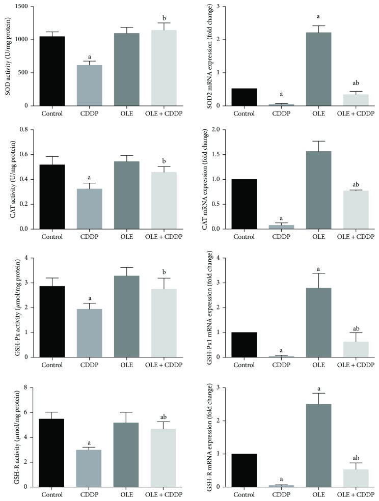
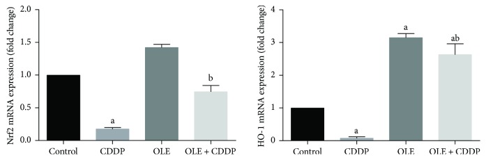
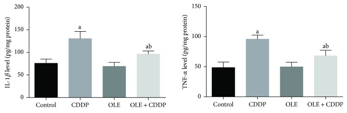
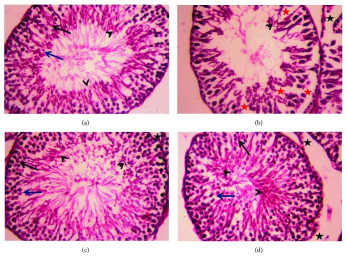
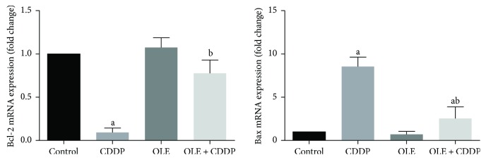
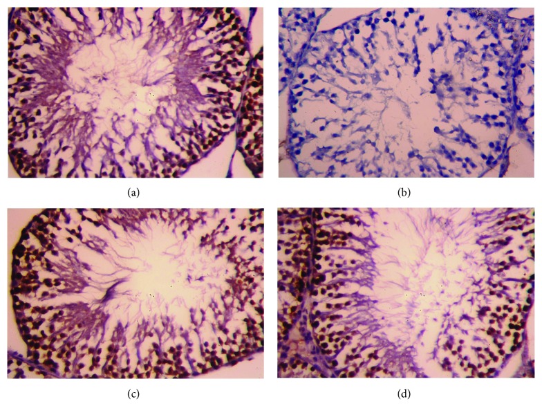
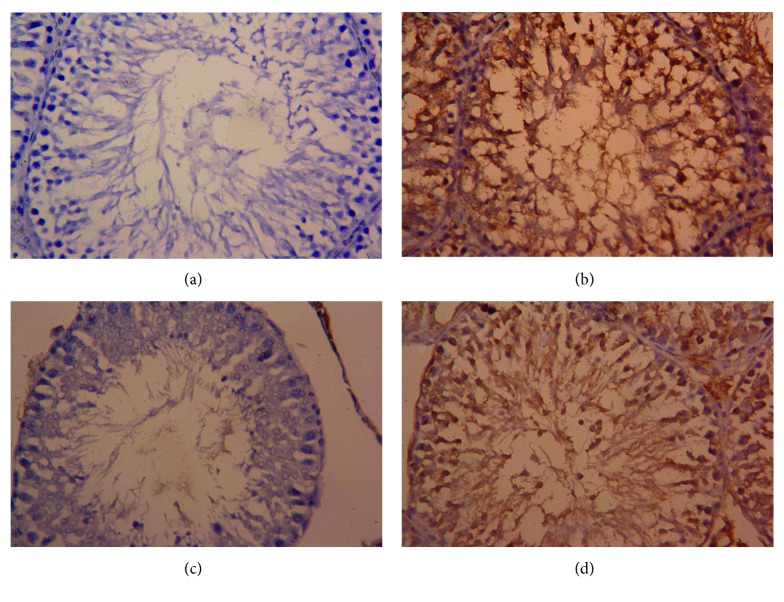
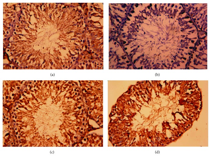
In the present investigation, the effect of olive leaf extract (OLE) on testicular damage induced in rats by an intraperitoneal injection of cisplatin (cis-diamminedichloroplatinum (CDDP)) at a dose of 5 mg/kg was tested. Rats were randomly divided into 4 groups: control, CDDP, OLE, and OLE + CDDP. After 5 days of CDDP treatment, body and testicular weights, histopathological alteration, and serum male sex hormone levels were determined. In addition to the biochemical and immunohistochemical changes in the testes, CDDP caused the disorganization of germinal epithelium and apoptosis by inducing Bax and inhibiting Bcl-2 protein expression. Testicular weights, catalase, serum testosterone, testicular enzymatic (including glutathione peroxidase, glutathione reductase, and superoxide dismutase) along with nonenzymatic (glutathione) antioxidants, and levels of luteinizing and follicle-stimulating hormones were significantly reduced in addition to a significant increase in testicular malondialdehyde and nitrite/nitrate levels when compared with the control group. OLE treatment markedly attenuated both biochemical and histopathological changes. The reproductive beneficial effects of OLE were mediated, at least partly, by inducing the nuclear factor erythroid 2-related factor 2 (Nrf2)/heme oxygenase 1 (HO-1) pathway.


Figures

Figure 1
Figure 1
Potential effects of olive leaf extract (OLE) treatment on body weight, testis weight, and relative testicular weight in rats treated with cisplatin (CDDP). All data are expressed as the mean ± SEM (n = 7). aSignificant change from the control group at p < 0.05; bsignificant change from the CDDP group at p < 0.05 using Tukey's post hoc test.
Figure 2
Figure 2
Effects of olive leaf extract (OLE) treatment on testosterone, luteinizing hormone, and follicle-stimulating hormone levels in the serum of rats treated with cisplatin (CDDP). All data are expressed as the mean ± SEM (n = 7). aSignificant change from the control group at p < 0.05; bsignificant change from the CDDP group at p < 0.05 using Tukey's post hoc test.
Figure 3
Figure 3
Effects of olive leaf extract (OLE) treatment on malondialdehyde (MDA), nitrite/nitrate, and glutathione (GSH) content in the testis of rats treated with cisplatin (CDDP). All data are expressed as the mean ± SEM (n = 7). aSignificant change from the control group at p < 0.05; bsignificant change from the CDDP group at p < 0.05 using Tukey's post hoc test.
Figure 4
Figure 4
Effects of olive leaf extract (OLE) treatment on superoxide dismutase (SOD), catalase (CAT), glutathione peroxidase (GSH-Px), and glutathione reductase (GSH-R) activities and their corresponding mRNA expression in the testis of rats treated with cisplatin (CDDP). Data of antioxidant enzyme activities are expressed as the mean ± SEM (n = 7), whereas mRNA expression data are expressed as the mean ± SEM of triplicate assays, normalized to the GAPDH mRNA level, and shown as fold change (in log2 scale) relative to the control mRNA levels. aSignificant change from the control group at p < 0.05; bsignificant change from the CDDP group at p < 0.05 using Tukey's post hoc test.
Figure 5
Figure 5
Effects of olive leaf extract (OLE) treatment on nuclear factor erythroid 2-related factor (Nrf2) and heme oxygenase-1 (HO-1) mRNA expression in the testis of rats treated with cisplatin (CDDP). Data of the mRNA expression are expressed as the mean ± SEM of triplicate assays, normalized to the GAPDH mRNA level, and shown as fold change (in log2 scale) relative to the control mRNA levels. aSignificant change from the control group at p < 0.05; bsignificant change from the CDDP group at p < 0.05 using Tukey's post hoc test.
Figure 6
Figure 6
Effects of olive leaf extract (OLE) treatment on TNF-α and IL-1β levels in the testis of rats treated with cisplatin (CDDP). All data are expressed as the mean ± SEM (n = 7). aSignificant change from the control group at p < 0.05; bsignificant change from the CDDP group at p < 0.05 using Tukey's post hoc test.
Figure 7
Figure 7
Light micrographs of testicular tissues of rats treated with olive leaf extract (OLE) and cisplatin (CDDP). (a) Photomicrograph of the testicular tissue of the control group showing healthy seminiferous tubules at all stages of spermatogenic cells (primary spermatocyte “black arrow” and spermatids “blue arrow”) and the interstitial cells with Leydig cells (black star) filling the space between the seminiferous tubules. (b) Photomicrograph of the testicular tissue of rats treated with CDDP showing degenerative alterations (red star) in spermatogenic cells and the detachment of the spermatogenic epithelium. (c) Photomicrograph of the testicular tissue of rats treated with OLE alone showing a healthy histological structure. (d) Photomicrograph of the testicular tissue of rats treated with OLE and CDDP showing a recovery of spermatogenic epithelium in most seminiferous tubules. Sections were stained with hematoxylin and eosin (400x).
Figure 8
Figure 8
Effects of olive leaf extract (OLE) treatment on Bcl-2 and Bax mRNA expression in the testis of rats treated with cisplatin (CDDP). Data of the mRNA expression are expressed as the mean ± SEM of triplicate assays, normalized to the GAPDH mRNA level, and shown as the fold change (in log2 scale) relative to the control mRNA levels. aSignificant change from the control group at p < 0.05; bsignificant change from the CDDP group at p < 0.05 using Tukey's post hoc test.
Figure 9
Figure 9
Testicular expression of Bcl-2 protein was detected using immunohistochemical staining in (a) control, (b) cisplatin (CDDP), (c) olive leaf extract (OLE), and (d) OLE + CDDP groups. In the control and OLE groups, Bcl-2-positive brown-stained cells were moderately to strongly immunostained. However, many testicular cells were weakly stained with brown color due to CDDP. In the OLE + CDDP group, the number of Bcl-2-positive cells was markedly increased. (400x).
Figure 10
Figure 10
Testicular expression of Bax protein was detected using immunohistochemical staining in (a) control, (b) cisplatin (CDDP), (c) olive leaf extract (OLE), and (d) OLE + CDDP groups. In the control and OLE groups, Bax-positive brown-stained cells were sparse and weakly immunostained. However, many testicular cells exhibited apoptosis and were stained brown (Bax positive) due to CDDP. In the OLE + CDDP group, the number of Bax-positive cells was markedly increased. (400x).
Figure 11
Figure 11
Testicular expression of proliferating cell nuclear antigen (PCNA) protein was detected using immunohistochemical staining in (a) control, (b) cisplatin (CDDP), (c) olive leaf extract (OLE), and (d) OLE + CDDP groups. In the control and OLE groups, PCNA-positive brown-stained cells were moderately to strongly immunostained. However, many testicular cells were weakly stained with brown color due to CDDP. In the OLE + CDDP group, the number of PCNA-positive cells was markedly increased. (400x).

References

    1. Abdel Moneim A. E. Azadirachta indica attenuates cisplatin-induced neurotoxicity in rats. Indian Journal of Pharmacology. 2014;46(3):316–321. doi: 10.4103/0253-7613.132182. - DOI - PMC - PubMed
    1. Sherif I. O., Abdel-Aziz A., Sarhan O. M. Cisplatin-induced testicular toxicity in rats: the protective effect of arjunolic acid. Journal of Biochemical and Molecular Toxicology. 2014;28(11):515–521. doi: 10.1002/jbt.21593. - DOI - PubMed
    1. Reddy K. P., Madhu P., Reddy P. S. Protective effects of resveratrol against cisplatin-induced testicular and epididymal toxicity in rats. Food and Chemical Toxicology. 2016;91:65–72. doi: 10.1016/j.fct.2016.02.017. - DOI - PubMed
    1. Fallahzadeh A. R., Rezaei Z., Rahimi H. R., et al. Evaluation of the effect of pentoxifylline on cisplatin-induced testicular toxicity in rats. Toxicology Research. 2017;33(3):255–263. doi: 10.5487/TR.2017.33.3.255. - DOI - PMC - PubMed
    1. Abdel Moneim A. E., Othman M. S., Aref A. M. Azadirachta indica attenuates cisplatin-induced nephrotoxicity and oxidative stress. BioMed Research International. 2014;2014:11. doi: 10.1155/2014/647131.647131 - DOI - PMC - PubMed

MeSH terms

LinkOut - more resources