

Exploring the villus
Abstract
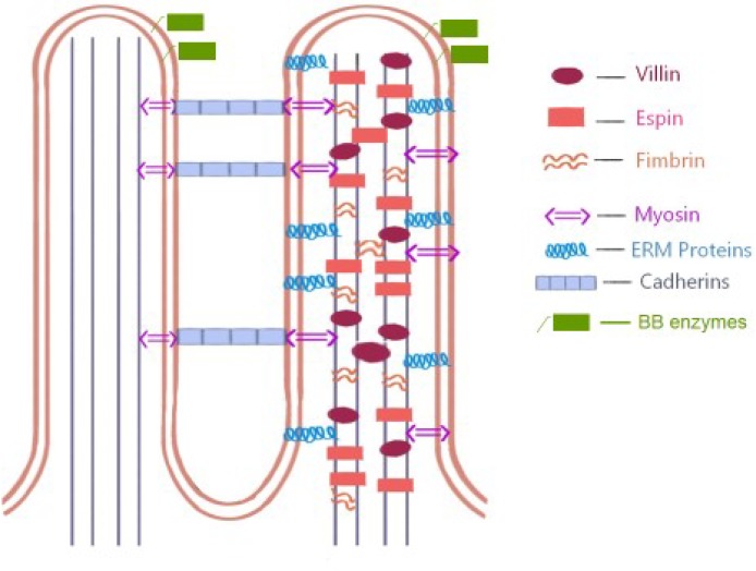
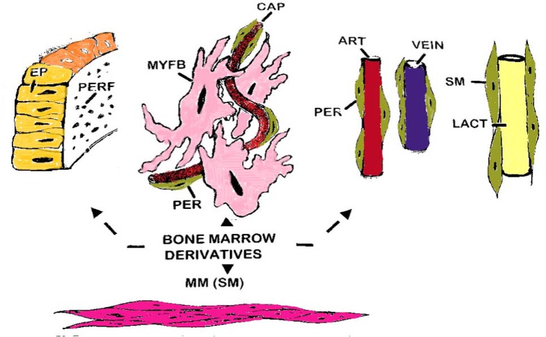
The small intestinal villus and its associated epithelium includes enterocytes as the main cell type and differentiated goblet and argentaffin cells, while the invaginated crypt epithelium is the site of cell division and hence the origin of all epithelial components. Enterocytes form a cohesive monolayer which acts both as a permeability barrier between lumen and the interior, and an important gateway for nutrient digestion, absorption and transport. Differentiation and polarisation of enterocytes depends on cytoskeletal proteins that control cell shape and maintain functionally specialised membrane domains; extracellular matrix (ECM) receptors; channels and transporters regulating ion/solute transfer across the cell. The mesenchymally-derived basement membrane dynamically controls morphogenesis, cell differentiation and polarity, while also providing the structural basis for villi, crypts and the microvasculature of the lamina propria so that tissue morphology, crucially, is preserved in the absence of epithelium. Mucosal re-organisation requires immense cooperation between all elements within the lamina, including marked revisions of the microvasculature and extensive alterations to all basement membranes providing support for endodermal and mesenchymal components. In this context, subepithelial myofibroblasts fulfil important regulatory activities in terms of tissue morphogenesis; remodelling; control of epithelial cell development, polarity and functional attributes; and an intimate involvement in repair, inflammation and fibrosis. This paper reviews the main structural and functional aspects of the villus, including the epithelium and its outer glycocalyx and microvillous border; and subjacent to the epithelium, the basement membrane with its attached web of myo-fibroblasts together with the lamina propria core of the villi, and its microvasculature and lacteals. Finally, some comments on the rapidity with which the overall structure of the villi changes in their response to both external, and internal, influences.
Keywords: Epithelium; Permeability; Small intestinal villus.
Figures
References
-
- Cruveilhier J, See M, editors. Traite d'Anatomie. 5th Ed. Paris: Asselin; 1874.
-
- Bichat X, editor. Traite des Membranes en General et de Diverses Membranes en Particuleur. Paris: ReInk Books; 1800.
-
- Henle FGJ. Symbolae ad Anatomian Villorum Intestinalium Imprimis Eorum et Vasorum Lacteorum. Berolini; 1837.
-
- Bowman W. "Mucous Membrane". In: Todd, RB, editor. Cyclopaedia of Anatomy & Physiology. Vol 3. London: Sherwood, Gilbert & Piper; 1847.
-
- Cheng H, Leblond CP. Origin, differentiation and renewal of the four main intestinal types in the mouse small intestine. Amer J Anat. 1974;141:461–562. - PubMed
Publication types
LinkOut - more resources
Full Text Sources