

doi: 10.1016/j.eats.2018.02.016.
eCollection 2018 Jun.
Is It Safe to Perform an Early Arthroscopy After a Traumatic Hip Dislocation With an Associated Pelvic Ring Injury? Report of Our Technique
Affiliations
- PMID: 30013910
- PMCID: PMC6020009
- DOI: 10.1016/j.eats.2018.02.016
Item in Clipboard
Is It Safe to Perform an Early Arthroscopy After a Traumatic Hip Dislocation With an Associated Pelvic Ring Injury? Report of Our Technique
Arthrosc Tech.
.
Abstract
Hip arthroscopy is useful in the treatment of several intra-articular pathologies, however, its use in high-energy hip and pelvis injuries raises concerns about fluid extravasion and stability of the pelvic ring. We present our arthroscopic surgical technique (initial access to the peripheral compartment) to remove intra-articular loose bodies and treat associated lesions, as well as our concerns with the technique, in case of a traumatic hip dislocation associated with a contralateral pelvic ring injury.
Figures
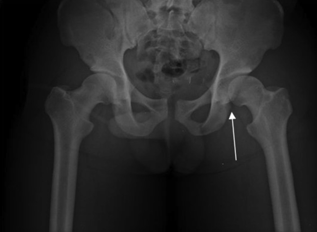
Pelvis radiograph showing the anteroinferior left hip dislocation and the contralateral wing of ilium fracture.
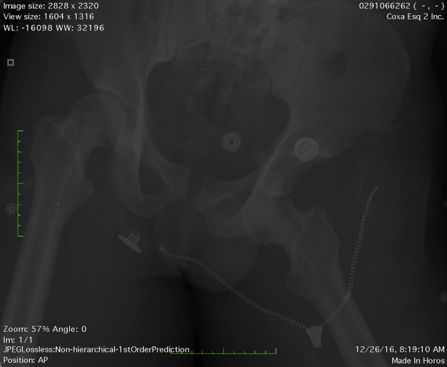
Anteroposterior pelvis radiograph in dorsal decubitus after reduction of the dislocation showing a nonconcentric reduction of the left hip, right iliac fracture, widening, and slight translation of the pubic symphysis. The arrow shows increased medial joint space.
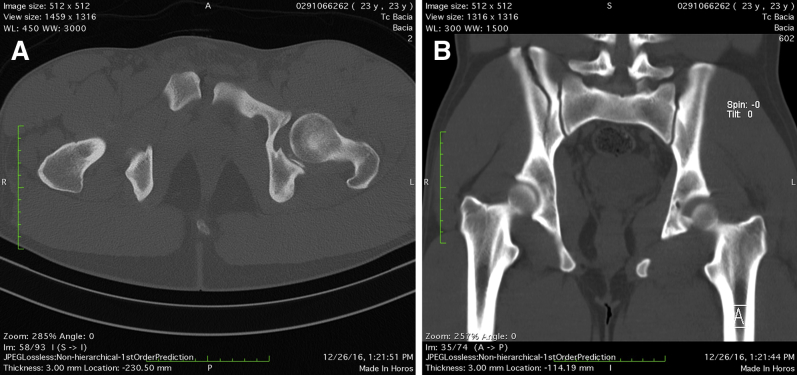
Pelvic computed tomography after reduction. (A) Axial reconstruction showing widening and anterior translation of the right pubic bone. (B) Coronal reconstruction showing a fracture of the right iliac wing without involvement of the sacroiliac joint (this fracture is complete). Both images show a nonconcentric reduction and the presence of intra-articular posteroinferior bone fragments interposed between the femoral head and acetabulum.
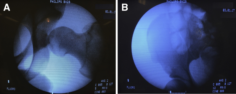
Preoperative fluoroscopy during traction test of the left hip (A). No fracture deviation was shown (B).
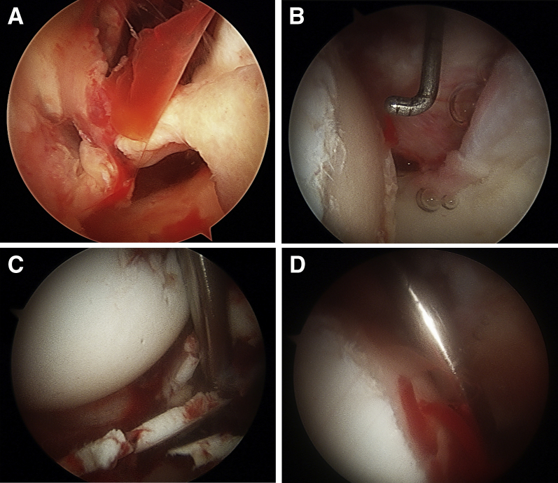
Left hip arthroscopy. (A) Intra-articular hematoma and rupture of the anteroinferior capsule; (B) chondral lesion of the femoral head with subchondral bone exposure at the central compartment, (C) intra-articular chondral fragments, and (D) microfractures on the femoral head, which continued to bleed.
References
-
- Phillips B.B., Mihalko M.J. Arthroscopy of the lower extremity. In: Azar F.M., Beaty J.H., Canale S.T., editors. Campbell's operative orthopaedics. Elsevier; Philadelphia: 2017. pp. 2486–2566.
-
- Egol K.A., Koval K.J., Zuckerman J.D. Wolters Kluwer Health; Philadelphia: 2015. Handbook of fractures.
-
- Kocher M.S., Frank J.S., Nasreddine A.Y. Intra-abdominal fluid extravasation during hip arthroscopy: a survey of the MAHORN group. Arthroscopy. 2012;28:1654–1660. - PubMed
-
- Kindler M., Halfmann B., Schoepp C., Rixen D. Hüftgelenkarthroskopie nach Luxatio obturatoria. Der Unfallchirurg. 2016;119:62–68. - PubMed
LinkOut - more resources
Full Text Sources
Other Literature Sources
