. 2018 Aug;24(8):1482-1489.
doi: 10.3201/eid2408.161888.

Susceptibility of Human Prion Protein to Conversion by Chronic Wasting Disease Prions

Susceptibility of Human Prion Protein to Conversion by Chronic Wasting Disease Prions

Marcelo A Barria et al. Emerg Infect Dis. 2018 Aug.

Abstract

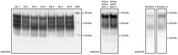
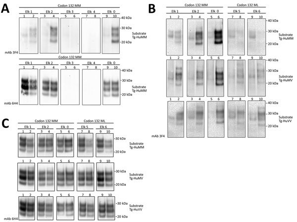
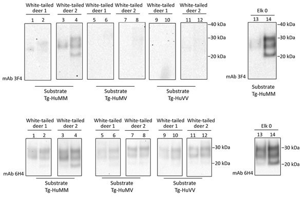
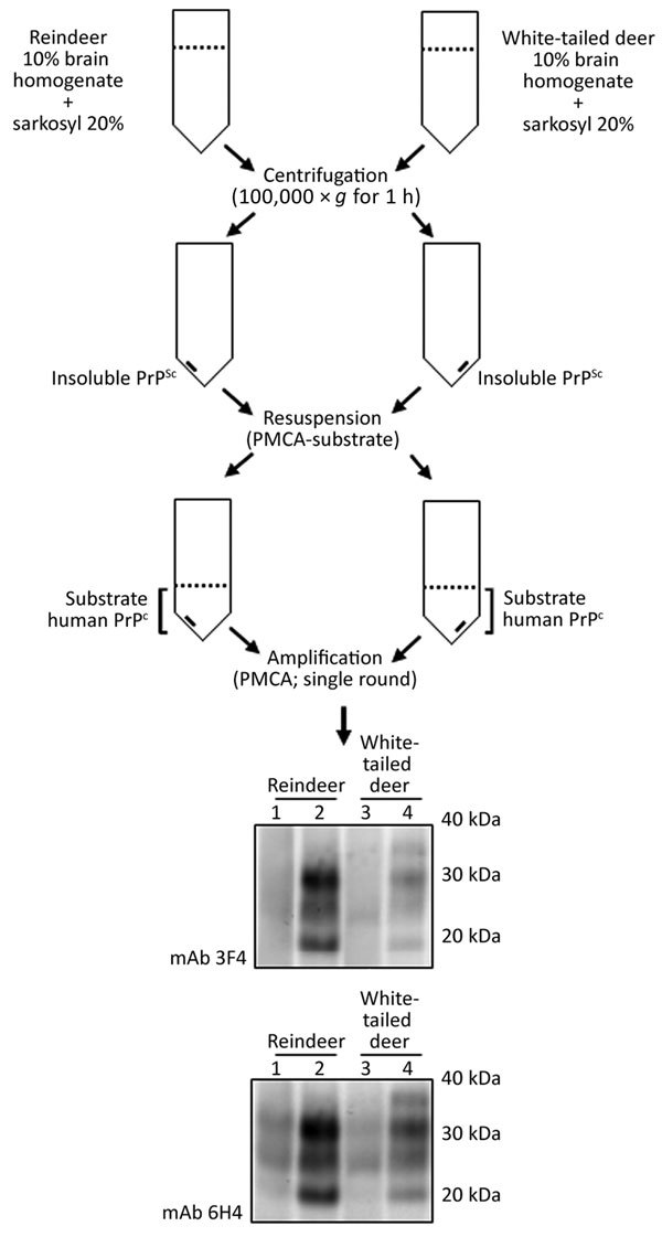
Chronic wasting disease (CWD) is a contagious and fatal neurodegenerative disease and a serious animal health issue for deer and elk in North America. The identification of the first cases of CWD among free-ranging reindeer and moose in Europe brings back into focus the unresolved issue of whether CWD can be zoonotic like bovine spongiform encephalopathy. We used a cell-free seeded protein misfolding assay to determine whether CWD prions from elk, white-tailed deer, and reindeer in North America can convert the human prion protein to the disease-associated form. We found that prions can convert, but the efficiency of conversion is affected by polymorphic variation in the cervid and human prion protein genes. In view of the similarity of reindeer, elk, and white-tailed deer in North America to reindeer, red deer, and roe deer, respectively, in Europe, a more comprehensive and thorough assessment of the zoonotic potential of CWD might be warranted.

Keywords: Europe; North America; chronic wasting disease; deer; elk; prion; protein misfolding cyclic amplification; reindeer; sheep; zoonoses.


Figures

Figure 1
Figure 1
Western blot analysis showing detectable levels of prion protein in the chronic wasting disease–affected cervid brain specimens used to evaluate the susceptibility of the human prion protein (PrP) to conversion by chronic wasting disease prions. We analyzed brain homogenate derived from elk, white-tailed deer, and reindeer specimens by using Western blot to evaluate levels of total PrP. We subjected 2 μL of each 10% brain homogenate sample to Western blot and assessed detection of total PrP by mAb 6H4. We performed 3 technical repeats with similar results; a representative Western blot is shown. Reference molecular markers have been included. mAb, monoclonal antibody.
Figure 2
Figure 2
Evaluation of the in vitro conversion of human prion protein (PrP) seeded with the misfolded, disease-associated prion protein form present in chronic wasting disease (CWD)–affected elk brain samples. Western blot analysis for PrP with odd and even number lanes showing reaction mixtures before and after protein misfolding cyclic amplification. A) We incubated 5 elk CWD specimens (elk 0–4) homozygous for Prnp codon 132 methionine (MM) in Tg-HuMM brain substrate (diluted 1:3) and subjected them to a single round of protein misfolding cyclic amplification followed by proteinase K digestion. We performed Western blot analysis by using the mAb 3F4 (for the detection of human protease-resistant prion protein [PrPres]) and 6H4 (detection of CWD PrPres and human PrPres). B) We used a panel of 3 humanized transgenic substrates (Tg-HuMM, Tg-Hu-MV, and Tg-HuVV) to evaluate the susceptibility of the human PrP to conversion. We assessed 3 CWD elk seeds of the132 MM genotype and 2 of the 132 methionine–leucine (ML) genotype. We detected conversion of the human PrP by CWD prions by using the mAb 3F4 after proteinase K digestion. C) We detected total PrPres by using Western blot with mAb 6H4. The elk specimen previously reported (15) is designated elk 0. We performed >5 repeats for the amplification of elk CWD 132 MM seeds and >3 for the 132 ML specimens with similar results. Reference molecular markers have been included. Molecular mass of electrophoretic markers is given. mAb, monoclonal antibody; Tg-HuMM, humanized transgenic PRNP codon 129 homozygous methionine; Tg-HuMV, humanized transgenic methionine/valine; Tg-HuVV, humanized transgenic valine/valine.
Figure 3
Figure 3
Evaluation of the in vitro conversion of human prion protein (PrP) seeded with the misfolded, disease-associated prion protein form present in chronic wasting disease (CWD)–affected white-tailed deer brain samples. We incubated 2 white-tailed deer CWD brain homogenates, derived from 2 affected animals (white-tailed deer 1 and 2), in a panel of 3 humanized transgenic substrates (Tg-HuMM, Tg-HuMV, and Tg-HuVV) and subjected them to a single round of protein misfolding cyclic amplification (PMCA) followed by proteinase K digestion. We diluted CWD brain homogenate 1:3 in PMCA substrate and performed Western blot analysis by using the mAb 3F4 (for the detection of human protease-resistant prion protein [PrPres]) and mAb 6H4 (for detection of CWD PrPres and human PrPres). We incorporated the elk specimen designated elk 0 as a control. We performed >3 repeats for the amplification white-tailed deer CWD 1 and 2 specimens with similar results. Reference molecular markers have been included. Molecular mass of electrophoretic markers is given. Odd and even number lanes show reaction mixtures before and after PMCA. mAb, monoclonal antibody; Tg-HuMM, humanized transgenic PRNP codon 129 homozygous methionine; Tg-Hu-MV, humanized transgenic methionine/valine; Tg-HuVV, humanized transgenic valine/valine.
Figure 4
Figure 4
Evaluation of the in vitro conversion of human prion protein (PrP) seeded with the misfolded, disease-associated prion protein form present in chronic wasting disease (CWD)–affected reindeer brain samples. We incubated 2 reindeer CWD specimens (reindeer 1 and 2) in a panel of 3 humanized transgenic substrates (Tg-HuMM, Tg-HuMV, and Tg-HuVV) and subjected them to a single round of protein misfolding cyclic amplification (PMCA). We diluted PMCA seeds 3 times in fresh PMCA substrate (dilution factor 1:3) and evaluated PMCA reactions for the presence of protease-resistant prion protein (PrPres) by proteinase K digestion. We performed Western blot analysis by using mAb 3F4 (for the detection of human PrPres) and mAb 6H4 (for detection of CWD PrPres and human PrPres). We incorporated the elk specimen designated elk 0 as a control. We performed >5 repeats for the amplification of reindeer 1 and 2 specimens. Reference molecular markers have been included. Molecular mass of electrophoretic markers is given. Odd and even number lanes show reaction mixtures before and after PMCA. mAb, monoclonal antibody; Tg-HuMM, humanized transgenic PRNP codon 129 homozygous methionine; Tg-Hu-MV, humanized transgenic methionine/valine; Tg-HuVV, humanized transgenic valine/valine.
Figure 5
Figure 5
Schematic representation of the partial purification of misfolded, disease-associated prion protein from chronic wasting disease (CWD)–affected deer brain specimens and its continued ability to seed the conversion of human prion protein (PrP) during protein misfolding cyclic amplification (PMCA) reactions. We normalized PrP, partially purified by detergent insolubility from reindeer and white-tailed deer CWD specimens, by using protease-resistant prion protein (PrPres) and subjected PrP to a single round of PMCA in humanized transgenic PRNP codon 129 homozygous methionine. We performed Western blot analysis by using mAb 3F4 (for detection of human PrPres) and mAb 6H4 (for detection of CWD PrPres and human PrPres). Molecular mass of electrophoretic markers is given. Odd and even number lanes show reaction mixtures before and after PMCA. mAb, monoclonal antibody; PMCA, protein misfolding cyclic amplification; PrPc, normal isoform of the prion protein; PrPsc, disease-associated isoform of the prion protein.

References

    1. Haley NJ, Hoover EA. Chronic wasting disease of cervids: current knowledge and future perspectives. Annu Rev Anim Biosci. 2015;3:305–25. 10.1146/annurev-animal-022114-111001 - DOI - PubMed
    1. Benestad SL, Mitchell G, Simmons M, Ytrehus B, Vikøren T. First case of chronic wasting disease in Europe in a Norwegian free-ranging reindeer. Vet Res (Faisalabad). 2016;47:88. 10.1186/s13567-016-0375-4 - DOI - PMC - PubMed
    1. Dagleish MP. Chronic wasting disease of deer—is the battle to keep Europe free already lost? Vet Rec. 2016;179:121–3. 10.1136/vr.i4165 - DOI - PubMed
    1. Head MW. Human prion diseases: molecular, cellular and population biology. Neuropathology. 2013;33:221–36. 10.1111/neup.12016 - DOI - PubMed
    1. Cassard H, Torres JM, Lacroux C, Douet JY, Benestad SL, Lantier F, et al. Evidence for zoonotic potential of ovine scrapie prions. Nat Commun. 2014;5:5821. 10.1038/ncomms6821 - DOI - PubMed

Publication types

LinkOut - more resources