. 2018 Sep;53(3):973-986.
doi: 10.3892/ijo.2018.4480. Epub 2018 Jul 12.

NLRP3 in human glioma is correlated with increased WHO grade, and regulates cellular proliferation, apoptosis and metastasis via epithelial-mesenchymal transition and the PTEN/AKT signaling pathway

Affiliations

NLRP3 in human glioma is correlated with increased WHO grade, and regulates cellular proliferation, apoptosis and metastasis via epithelial-mesenchymal transition and the PTEN/AKT signaling pathway

Xiao-Feng Yin et al. Int J Oncol. 2018 Sep.

Abstract

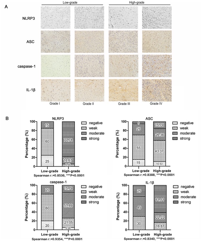
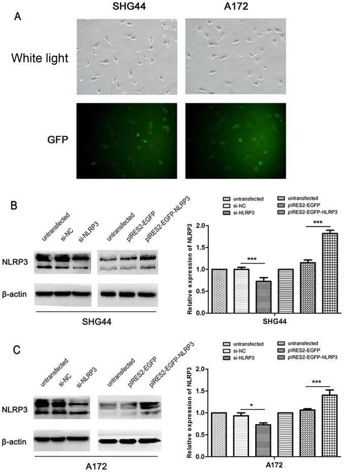
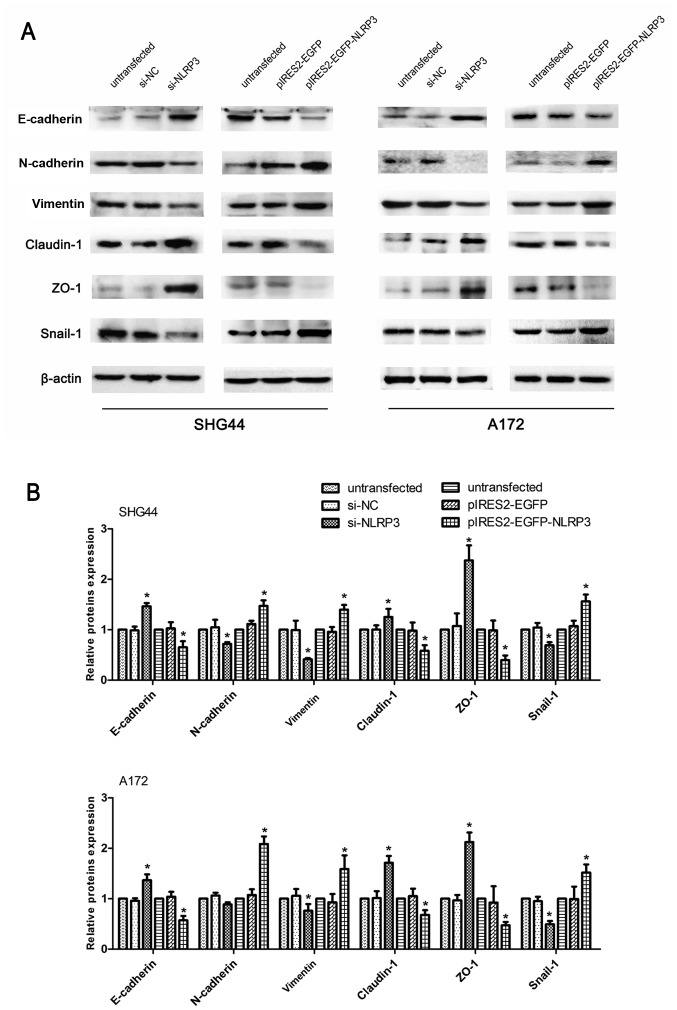
Glioma is the most prevalent and fatal primary tumor of the central nervous system in adults, while the development of effective therapeutic strategies in clinical practice remain a challenge. Nucleotide-binding domain leucine-rich family pyrin-containing 3 (NLRP3) has been reported to be associated with tumorigenesis and progression; however, its expression and function in human glioma remain unclear. The present study was designed to explore the biological role and potential mechanism of NLRP3 in human glioma. The results demonstrated that overexpression of NLRP3, apoptosis-associated speck-like protein containing a caspase-recruitment domain (ASC), caspase‑1 and interleukin (IL)‑1β protein in human glioma tissues were significantly correlated with higher World Health Organization grades. The in vitro biological experiments demonstrated that NLRP3 downregulation significantly inhibited the proliferation, migration and invasion, and promoted the apoptosis of SHG44 and A172 glioma cell lines. Furthermore, western blot assays revealed that the downregulation of NLRP3 significantly reduced the expression of ASC, caspase‑1 and IL‑1β protein. Furthermore, NLRP3 knockdown caused the inhibition of epithelial-mesenchymal transition (EMT), and inhibited the phosphorylation of AKT serine/threonine kinase (AKT) and phosphorylation of phosphatase and tensin homolog (PTEN). Consistently, the upregulation of NLRP3 significantly increased the expression of ASC, caspase‑1, IL‑1β and phosphorylated-PTEN, promoted proliferation, migration, invasion and EMT, inhibited apoptosis, and activated the AKT signaling pathway. The data of the present study indicate that NLRP3 affects human glioma progression and metastasis through multiple pathways, including EMT and PTEN/AKT signaling pathway regulation, enhanced inflammasome activation, and undefined inflammasome-independent mechanisms. Understanding the biological effects of NLRP3 in human glioma and the underlying mechanisms may offer novel insights for the development of glioma clinical therapeutic strategies.


Figures

Figure 1
Figure 1
NLRP3, ASC, caspase-1 and IL-1β tissue expression in low-grade glioma (WHO I and II) and high-grade glioma was analyzed by immunohisto-chemical staining. (A) Representative images are shown at magnification, ×100. (B) Statistical analyses were performed to determine whether the expression levels of NLRP3, ASC, caspase-1 and IL-1β in human glioma tissue correlated with histological grades. NLRP3, NLR family pyrin domain containing 3; ASC, apoptosis-associated speck-like protein containing a caspase-recruitment domain; IL, interleukin.
Figure 2
Figure 2
Identification of the efficiency of NLRP3 downregulation and overexpression in glioma cell lines, SHG44 and A172. (A) The transfection efficiency of plasmid vector was indicated using bright field and GFP (magnification, ×100); (B) Western blot analysis detected the downregulation and overexpression efficiency of NLRP3 at protein levels. The intensity of protein bands was measured and the values are presented as the mean ± standard deviation (n=3). *P<0.05 and ***P<0.001. NLRP3, NLR family pyrin domain containing 3; GFP, green fluorescent protein; NC, negative control; EGFP, enhanced green fluorescent protein.
Figure 3
Figure 3
Facilitative effect of NLRP3 on the proliferation of SHG44 and A172 glioma cells. (A) The effect of NLRP3 on glioma cell viability was assessed using a colony formation assay. (B) A Cell Counting Kit-8 assay was used to measure cell viability in untransfected and transfected glioma cells. (C) An EdU assay was used to evaluate the proliferation rate of glioma cells. Representative images are shown at magnification, ×100 (A) and ×200 (C). Values are presented as the mean ± standard deviation (n=3). *P<0.05 and **P<0.01. NLRP3, NLR family pyrin domain containing 3; EdU, 5-ethynyl-2′-deoxyuridine; NC, negative control; EGFP, enhanced green fluorescent protein; OD, optical density.
Figure 4
Figure 4
Effect of NLRP3 on SHG44 and A172 glioma cell apoptosis. The apoptotic glioma cells labeled with Annexin V-APC and 7-AAD were detected by flow cytometry. Values of apoptosis rate were expressed as the mean ± standard deviation (n=3). **P<0.01 and***P<0.001. A representative apoptosis plot from three experiments performed is shown. NLRP3, NLR family pyrin domain containing 3; APC, allophycocyanin; 7-AAD, 7-Aminoactinomycin D; NC, negative control; EGFP, enhanced green fluorescent protein.
Figure 5
Figure 5
Effect of NLRP3 on promoting migration and invasion in SHG44 and A172 human glioma cells. (A) The effect of NLRP3 on glioma cells migratory ability was assessed using wound healing assays. The rate of wound closure was monitored by measuring the ratio of the distance of the wound at 0 and 24 h. (B) The effect of NLRP3 on glioma cells migratory ability was detected using Transwell migration assays. (C) The effects of NLRP3 on glioma cellular invasion ability were detected using Transwell invasion assays. Representative images of wound healing, Transwell migration and invasion assays from three independent experiments are presented. Values are expressed as the mean ± standard deviation (n=3). *P<0.05, **P<0.01 and ***P<0.001. NLRP3, NLR family pyrin domain containing 3; NC, negative control; EGFP, enhanced green fluorescent protein.
Figure 6
Figure 6
Regulatory effects of NLRP3 on intracellular inflammasome activation in glioma cells. (A) The effect of NLRP3 on altering the expression of molecules involved in the construction of inflammasome complex and its activation. The expression levels of ASC, caspase-1, pro-IL-1β and the molecules that indicate inflammasome activation, including cleaved-caspase-1 (p20) and mature IL-1β (17 kDa) were assessed by western blot analysis. (B) The intensity of protein bands was measured and the values as the mean ± standard deviation (n=3). *P<0.05. NLRP3, NLR family pyrin domain containing 3; ASC, apoptosis-associated speck-like protein containing a caspase-recruitment domain; IL, interleukin; NC, negative control; EGFP, enhanced green fluorescent protein.
Figure 7
Figure 7
Regulatory effects of NLRP3 on molecules involved in EMT in glioma cells. (A) The changes in EMT-associated proteins or transcriptional factors include E-cadherin, N-cadherin, vimentin, claudin-1, ZO-1 and snail-1 in glioma cell lines SHG44 and A172 were assessed by western blot analysis. (B) The intensity of protein bands was measured and the values are presented as the mean ± standard deviation (n=3). *P<0.05. NLRP3, NLR family pyrin domain containing 3; NC, negative control; EGFP, enhanced green fluorescent protein; EMT, epithelial-mesenchymal transition; ZO-1, zonulaoccluden-1.
Figure 8
Figure 8
Regulatory effects of NLRP3 on the PTEN/AKT signaling pathway in glioma cells. (A) Western blotting was used to determine the level of proteins associated with PTEN/AKT signaling pathway, including total AKT, p-AKT (Ser473), p-AKT (Thr308), GSK3β, p-GSK3β (Ser9), and its regulators p-PTEN (Ser380) and total PTEN. (B) The intensity of protein bands was measured and the values are presented as the mean ± standard deviation (n=3). *P<0.05. NLRP3, NLR family pyrin domain containing 3; NC, negative control; EGFP, enhanced green fluorescent protein; AKT serine/threonine kinase; GSK3β, glycogen synthase kinase-3β; PTEN, phosphatase and tensin homolog.

References

    1. Wen PY, Reardon DA. Neuro-oncology in 2015: Progress in glioma diagnosis, classification and treatment. Nat Rev Neurol. 2016;12:69–70. doi: 10.1038/nrneurol.2015.242. - DOI - PubMed
    1. Mao K, Lei D, Zhang H, You C. MicroRNA-485 inhibits malignant biological behaviour of glioblastoma cells by directly targeting PAK4. Int J Oncol. 2017;51:1521–1532. doi: 10.3892/ijo.2017.4122. - DOI - PubMed
    1. Omuro A, DeAngelis LM. Glioblastoma and other malignant gliomas: A clinical review. JAMA. 2013;310:1842–1850. doi: 10.1001/jama.2013.280319. - DOI - PubMed
    1. Olar A, Sulman EP. Molecular markers in low-grade glioma-toward tumor reclassification. Semin Radiat Oncol. 2015;25:155–163. doi: 10.1016/j.semradonc.2015.02.006. - DOI - PMC - PubMed
    1. Christofides A, Kosmopoulos M, Piperi C. Pathophysiological mechanisms regulated by cytokines in gliomas. Cytokine. 2015;71:377–384. doi: 10.1016/j.cyto.2014.09.008. - DOI - PubMed

MeSH terms