Case Definition of Chronic Pulmonary Aspergillosis in Resource-Constrained Settings

David W Denning et al. Emerg Infect Dis. 2018 Aug.

Abstract

Chronic pulmonary aspergillosis (CPA) is a recognized complication of pulmonary tuberculosis (TB). In 2015, the World Health Organization reported 2.2 million new cases of nonbacteriologically confirmed pulmonary TB; some of these patients probably had undiagnosed CPA. In October 2016, the Global Action Fund for Fungal Infections convened an international expert panel to develop a case definition of CPA for resource-constrained settings. This panel defined CPA as illness for >3 months and all of the following: 1) weight loss, persistent cough, and/or hemoptysis; 2) chest images showing progressive cavitary infiltrates and/or a fungal ball and/or pericavitary fibrosis or infiltrates or pleural thickening; and 3) a positive Aspergillus IgG assay result or other evidence of Aspergillus infection. The proposed definition will facilitate advancements in research, practice, and policy in lower- and middle-income countries as well as in resource-constrained settings.

Keywords: Aspergillus; Tuberculosis; antibody; aspergilloma; developing countries; fungi; imaging; resource-constrained settings; tuberculosis and other mycobacteria.


Figures

Figure 1
Figure 1
A) Chest radiograph showing aspergilloma (fungal ball) in the left upper lung lobe. B) Axial computed tomography image shows increased density in an irregular left apical cavity, a sequela of pulmonary tuberculosis, consistent with an aspergilloma.
Figure 2
Figure 2
Computed tomography images of a patient with chronic pulmonary aspergillosis. A) Left upper lung lobe thick-walled cavity, showing associated pleural thickening. B) Same patient several months later, demonstrating progression of cavitation with increased pericavitary consolidation and formation of a fungal ball within the cavity. Aspergilloma formation is a late feature of chronic pulmonary aspergillosis.
Figure 3
Figure 3
Computed tomography images showing early features of fungal ball formation in pulmonary cavities. A) Two left lower lung lobe posterior thick-walled cavities, 1 with a fluid level. B) Two right apical cavities, the larger with an irregular interior wall, most consistent with fungal growth. C) Left apex replaced by an irregular thick-walled cavity with multiple areas of fungal growth on the interior surface of the cavity. D) Substantial volume loss in the right upper lobe with replacement by a small anterior cavity and larger crescent-shaped cavity with both pleural thickening and fat indrawing along the pleural surface posteriorly. The cavity shows marked irregularity consistent with fungal growth. E) A right upper lobe thin-walled cavity containing 2 areas of fungal growth, 1 of which has detached from the wall as a thick mat of mycelial growth with a larger lump in the cavity interior. F) Multiple cavities in both upper lobes, with wall irregularity in the left upper lobe cavity consistent with surface fugal growth. The right upper lobe cavity shows pleural thickening and indrawing of fat posteriorly.
Figure 4
Figure 4
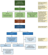
Diagnostic algorithm incorporating the chest radiographic appearance and results of rapid TB investigations with the case definition of CPA. ABPA, allergic bronchopulmonary aspergillosis; CPA, chronic pulmonary aspergillosis; CT, computed tomography; NTM, nontuberculous mycobacteria; TB, tuberculosis. GeneXpert, http://www.cepheid.com/us/cepheid-solutions/systems/genexpert-systems/genexpert-iv

References

    1. Goodwin RA Jr, Owens FT, Snell JD, Hubbard WW, Buchanan RD, Terry RT, et al. Chronic pulmonary histoplasmosis. Medicine (Baltimore). 1976;55:413–52. 10.1097/00005792-197611000-00001 - DOI - PubMed
    1. Shah A, Panjabi C. Allergic bronchopulmonary aspergillosis: a review of a disease with a worldwide distribution. J Asthma. 2002;39:273–89. 10.1081/JAS-120002284 - DOI - PubMed
    1. Chong S, Lee KS, Yi CA, Chung MJ, Kim TS, Han J. Pulmonary fungal infection: imaging findings in immunocompetent and immunocompromised patients. Eur J Radiol. 2006;59:371–83. 10.1016/j.ejrad.2006.04.017 - DOI - PubMed
    1. Chu HQ, Li B, Zhao L, Huang DD, Zhang ZM, Xu JF, et al. Chest imaging comparison between non-tuberculous and tuberculosis mycobacteria in sputum acid fast bacilli smear-positive patients. Eur Rev Med Pharmacol Sci. 2015;19:2429–39. - PubMed
    1. Chiang CY, Yu MC, Yang SL, Yen MY, Bai KJ. Surveillance of tuberculosis in Taipei: the influence of nontuberculous mycobacteria. PLoS One. 2015;10:e0142324. 10.1371/journal.pone.0142324 - DOI - PMC - PubMed

Publication types