. 2018 Nov:65:80-90.
doi: 10.1016/j.meegid.2018.07.016. Epub 2018 Jul 19.

Analysis of genetic polymorphisms and tropism in East African Leishmania donovani by Amplified Fragment Length Polymorphism and kDNA minicircle sequencing

Affiliations

Analysis of genetic polymorphisms and tropism in East African Leishmania donovani by Amplified Fragment Length Polymorphism and kDNA minicircle sequencing

Hanan T Jaber et al. Infect Genet Evol. 2018 Nov.

Abstract

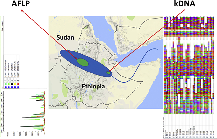
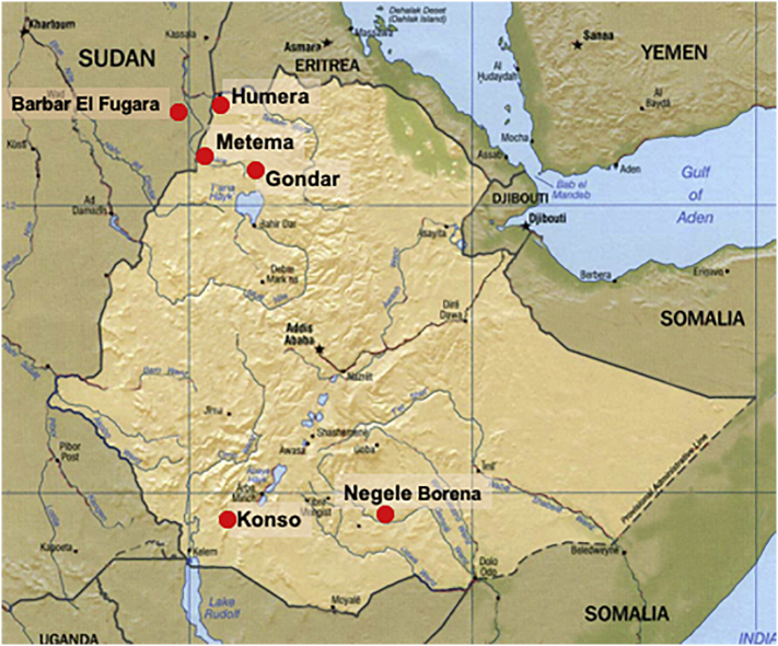
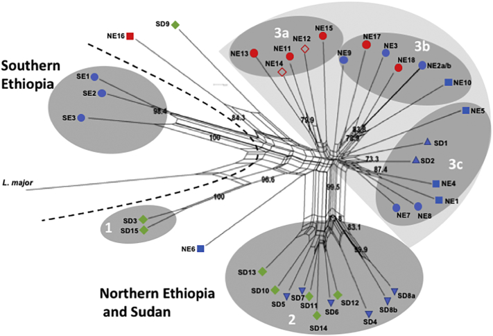
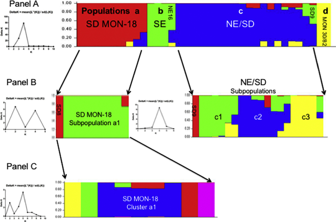
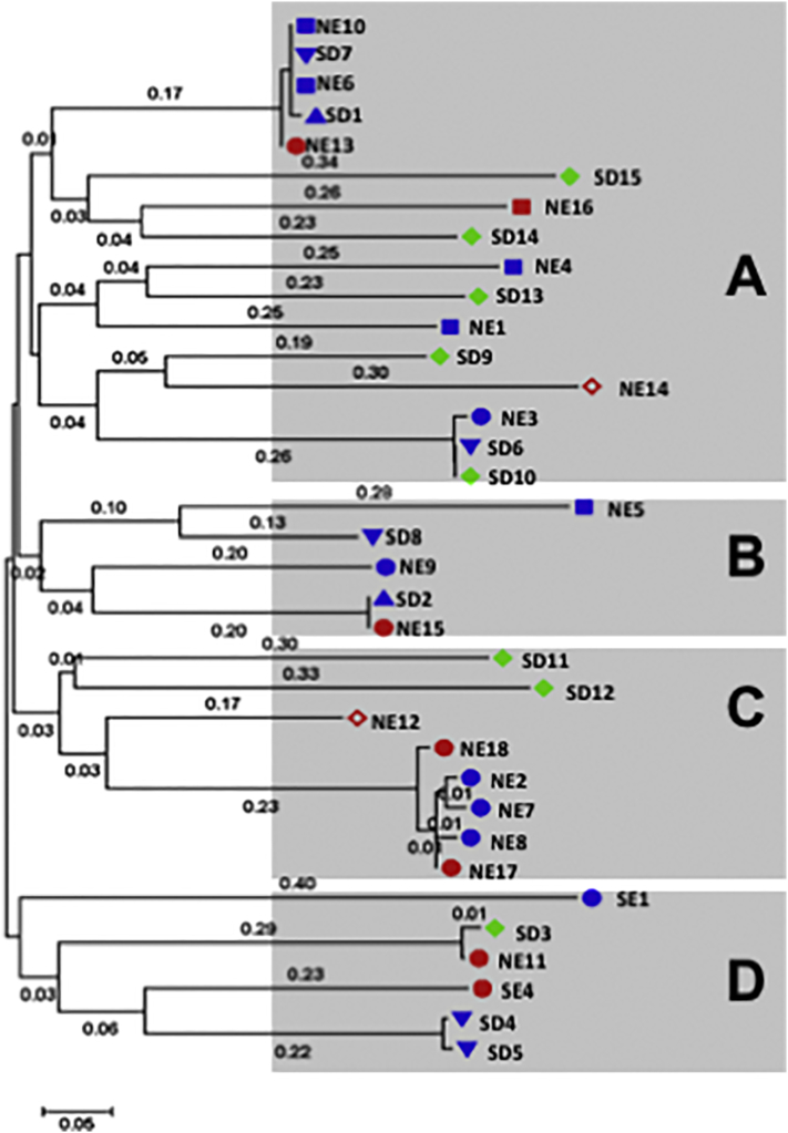
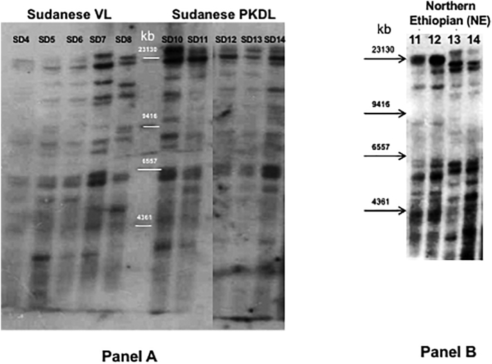
Visceral leishmaniasis (VL), the most severe form of leishmaniasis, is caused by Leishmania donovani. In addition to fatal VL, these parasites also cause skin diseases in immune-competent and -suppressed people, post-kala azar dermal leishmaniasis (PKDL) and HIV/VL co-infections, respectively. Genetic polymorphism in 36 Ethiopian and Sudanese L. donovani strains from VL, PKDL and HIV/VL patients was examined using Amplified Fragment Length Polymorphism (AFLP), kDNA minicircle sequencing and Southern blotting. Strains were isolated from different patient tissues: in VL from lymph node, spleen or bone marrow; and in HIV/VL from skin, spleen or bone marrow. When VL and PKDL strains from the same region in Sudan were examined by Southern blotting using a DNA probe to the L. donovani 28S rRNA gene only minor differences were observed. kDNA sequence analysis distributed the strains in no particular order among four clusters (A - D), while AFLP analysis grouped the strains according to geographical origin into two major clades, Southern Ethiopia (SE) and Sudan/Northern Ethiopia (SD/NE). Strains in the latter clade were further divided into subpopulations by zymodeme, geography and year of isolation, but not by clinical symptoms. However, skin isolates showed significantly (p < 0.0001) fewer polymorphic AFLP fragments (average 10 strains = 348.6 ± 8.1) than VL strains (average 26 strains = 383.5 ± 3.8).

Keywords: Amplified Fragment Length Polymorphism; Genetic polymorphism; HIV-visceral leishmaniasis co-infections; Leishmania donovani; Post-kala azar dermal leishmaniasis; kDNA minicircle sequence.


Figures

Unlabelled Image
Graphical abstract
Fig. 1
Fig. 1
Map indicating the sites where the Leishmania donovani strains examined in this study were isolated in Sudan and Ethiopia. This figure was adapted from Gelanew et al., 2010b.
Fig. 2
Fig. 2
Neighbor-net splits graph derived from combined analysis of AFLP using six selective primers. Results were analyzed using optiFLP (Arthofer et al., 2011) and tinyFLP (Arthofer, 2010), and the binary matrices concatenated using tinyCAT (Arthofer, 2010). The tree above was built using Neighbor-net analysis of the concatenated binary matrix with Splitstree 4 (Huson and Bryant, 2006). The numbers above each branch indicate the bootstrap support in percentage from 1000 replicates. SD1 and SD2 are reference strains from Sudan that were isolated in 1943 and 1962, respectively. Strains NE2a and NE2b, as well as SD8a and SD8b, are technical duplicates. Selective PCRs for these strains were run in duplicate using the same pre-selective PCR product. Strain origin: Northern Ethiopia (NE) groups 3a, 3b and 3c, Southern Ethiopia (SE) group1, and Sudan (SD) groups 2 and 3c. Clinical presentation: VL – blue, HIV/VL co-infection – red, PKDL – green. Tissue source: skin isolates – both full and empty diamonds, spleen isolates - circles, lymph node -inverted triangle, bone marrow isolates - squares, and no information - pyramid. Leishmania major was included as an outlier. (For interpretation of the references to colour in this figure legend, the reader is referred to the web version of this article.)
Fig. 3
Fig. 3
Population analysis of the 36 east African Leishmania donovani strains as determined by STRUCTURE (Pritchard et al., 2000) based on AFLP using six selective primers. A single vertical bar represents each strain with the different colors indicating to the fraction of genotype attributed by each population (Q). A single colour indicates that a strain belongs to only one genetic group. Population parameters were estimated by 10 Markov Chain Monte Carlo iterations after a burn-in of 100,000 simulations. Panel A. At K = 4 the strains group into four distinct populations which was confirmed by ∆K analysis, plot on left, using Structure Harvester (Earl and Vonholdt, 2012): a) SD MON-18 - all 10 Sudanese MON-18 strains; b) SE - the southern Ethiopian strains plus one atypical northern Ethiopian (NE) HIV/VL bone marrow strain (NE16); c) NE/SD - 17/18 NE strains and three of the SD strains: PKDL strain SD9 (MON-267), SD1 and SD2; and d) MON30/82 –PKDL strains SD3 and SD15 with zymodemes MON-82 and MON-30, respectively. Panel B. Further analysis of SD MON-18 and NE/SD by STRUCTURE identified additional subgroups. Subpopulation a1 contains all of the SD MON-18 strains except SD8, while the NE/SD population was divided into four subpopulations: c1 - NE1, NE4, NE6 - NE8 and SD2; c2 - NE2, NE3, NE5, NE9, NE10, NE17, NE18 and SD1; c3 - NE11 - NE15; and a fourth group containing the PKDL isolate SD9 (zymodeme MON-267). Panel C. Further analysis of SD MON-18 subpopulation a1. Cluster a1 contains SD6 SD7, SD10, SD11 and SD12.
Fig. 4
Fig. 4
Number of AFLP alleles for skin and non-skin Leishmania strains. AFLP analysis using six selective primers was performed on 36 L. donovani and one L. major strains. The total number of polymorphic AFLP fragments was plotted according to tissue from which strain was cultured. Skin: 10 strains were isolated from the skin of northern Ethiopian HIV/VL and Sudanese PKDL patients. Non-skin: 26 strains were isolated from the spleen, bone marrow and lymph nodes of VL and HIV/VL patients from Sudan and Ethiopia. Skin isolates (average 10 strains = 348.6 ± 8.1), VL strains (average 26 strains = 383.5 ± 3.8). *unpaired t-test, p < 0.0001.
Fig. 5
Fig. 5
Phylogenetic tree of East African Leishmania donovani strains based on analysis of kDNA minicircle sequences. The sequences (GeneBank accession numbers KY950645 to KY950679) were aligned using Muscle algorithm with gaps, and the phylogenetic tree was built using the Maximum Likelihood method based on the Hasegawa-Kishino-Yano model (Hasegawa et al., 1985) using MEGA7 (Kumar et al., 2016) as described in Material and Methods. The tree is drawn to scale, with branch lengths measured in the number of substitutions per site (next to the branches). Strain origin: Northern (NE) or Southern Ethiopia (SE), Sudan (SD). Clinical presentation: VL – blue, HIV/VL co-infection – red, PKDL – green. Tissue source: skin isolates – full and empty diamonds, spleen isolates - circles, lymph node -inverted triangle, bone marrow isolates - squares, and no information - pyramid. Strains included in Group A – NE1, NE3, NE4, NE6, NE10, NE13, NE14, NE16, SD1, SD6, SD7, SD9, SD10, SD13 - SD15; Group B –NE5, NE9, NE15, SD2 and SD8; Group C – NE2, NE7, NE8, NE12, NE17, NE18, SD11 and SD12; Group D –NE11, SD3, SD4, SD5, SE1 and SE4. (For interpretation of the references to colour in this figure legend, the reader is referred to the web version of this article.)
Fig. S1
Fig. S1
Internal transcribed spacer 1 (ITS1)–PCR analysis of Sudanese Leishmania donovani strain SD9 (LEM3472). Panel A – Parasites were cloned (Garin et al., 2002), and ITS-1–PCR RFLP carried out (Schonian et al., 2003). Ethidium-bromide stained agarose gel showing the parent strain SD9 (MHOM/SD/97/LEM3472) with an extra band at 239 bp*, and another Sudanese strain SD13 (MHOM/SD/98/LEM3570) displaying typical HaeIII digestion patterns (189, 77 and 51 bp arrows). Panel B - Sequence alignment of ITS1–PCR region of the Sudanese PKDL strain MHOM/SD/97/LEM3472 (SD9). The PCR products were cloned into pGEM-T Easy and colonies picked for sequencing, and DNA sequences were compared using MultAlin (http://sacs.ucsf.edu/cgi-bin/multalin.py). Sequences examined: Genebank – SD9_database (accession number emb|AJ634370.1|), Parental strain – SD9_parental (this study), and SD9 amplicons subcloned into pGEM –T Easy - colony_5, _2, _14 and _18 (this study). In colony 14 a single nucleotide point mutation (C189A) is present eliminating the HaeIII restriction site (GGCC) resulting in an extra 239 bp product. Additional single nucleotide point mutations, colony 18 (T169C) and colony 2 (G318A) were noted suggesting that different ITS-1 sequences exist in the tandem repeats (Downing et al., 2011; Gelanew et al., 2010a) of the ITS1 regions between the SSU rRNA and the 5.8S genes on chromosome 27.
Fig. S2
Fig. S2
Southern blot of East African Leishmania donovani strains using a DNA probe (Ld28S) for the ζ and ε subunit regions of the 28S rRNA gene. Panel A: Sudanese VL or PKDL strains. Lanes 1–5: VL strains (SD4 – SD8); Lanes 7–11: PKDL strains (SD10 – SD14). Panel B; HIV/VL strains isolated from different organs, spleen (NE11 and NE13) or skin (NE12 and NE14), of patients GR363 (NE11 and NE12) and GR364 (NE13 and NE14).
Fig. S3
Fig. S3
Analysis by kDNA PCR-RFLP of HIV/VL strains isolated from different organs of the same patient. kDNA minicircles from three patients were amplified by PCR using the primer pair Uni21 and Lmj4 (Anders et al., 2002) and purified as described in Material and Methods. The purified products were digested overnight with HaeIII and separated by electrophoresis on 3% NuSieve 3:1 Agarose (Cambrex Bio Science Rockland, Inc.) gels. Lanes: Mr. - 100 kb marker; GR363 NE11 – spleen and NE12 – skin; GR364 NE13 – spleen and NE14 – skin; LDS373/08 NE15 – spleen and NE16 – bone marrow.

References

    1. Alonso D.P., Costa D.L., de Mendonca I.L., Costa C.H., Ribolla P.E. Heterogeneity of Leishmania infantum chagasi kinetoplast DNA in Teresina (Brazil) Am. J. Trop. Med. Hyg. 2010;82:819–821. - PMC - PubMed
    1. Alvar J., Bashaye S., Argaw D., Cruz I., Aparicio P., Kassa A., Orfanos G., Parreno F., Babaniyi O., Gudeta N., Canavate C., Bern C. Kala-azar outbreak in Libo Kemkem, Ethiopia: epidemiologic and parasitologic assessment. Am. J. Trop. Med. Hyg. 2007;77:275–282. - PubMed
    1. Alvar J., Aparicio P., Aseffa A., Den Boer M., Canavate C., Dedet J.P., Gradoni L., Ter Horst R., Lopez-Velez R., Moreno J. The relationship between leishmaniasis and AIDS: the second 10 years. Clin. Microbiol. Rev. 2008;21:334–359. - PMC - PubMed
    1. Alvar J., Vélez I.D., Bern C., Herrero M., Desjeux P., Cano J., Jannin J., den Boer M., Team W.L.C. Leishmaniasis worldwide and global estimates of its incidence. PLoS One. 2012;7 - PMC - PubMed
    1. Anders G., Eisenberger C.L., Jonas F., Greenblatt C.L. Distinguishing Leishmania tropica and Leishmania major in the Middle East using the polymerase chain reaction with kinetoplast DNA-specific primers. Trans. R. Soc. Trop. Med. Hyg. 2002;96:S87–S92. Suppl 1. - PubMed

Publication types

LinkOut - more resources