

Tumor thrombus in the large veins draining primary pelvic osteosarcoma on cross sectional imaging
- PMID: 30017298
- PMCID: PMC6056011
- DOI: 10.1016/j.ejrad.2018.05.021
Tumor thrombus in the large veins draining primary pelvic osteosarcoma on cross sectional imaging
Abstract
Purpose: To evaluate the frequency of tumor thrombus in the large veins draining primary pelvic osteosarcoma on early cross-sectional imaging studies and its effect on patient survival.
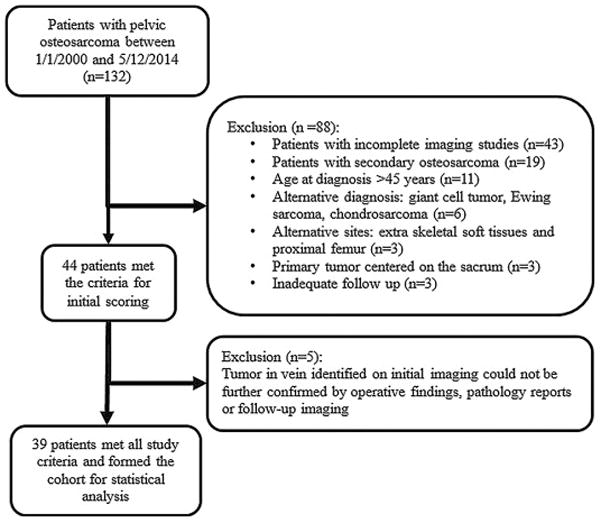
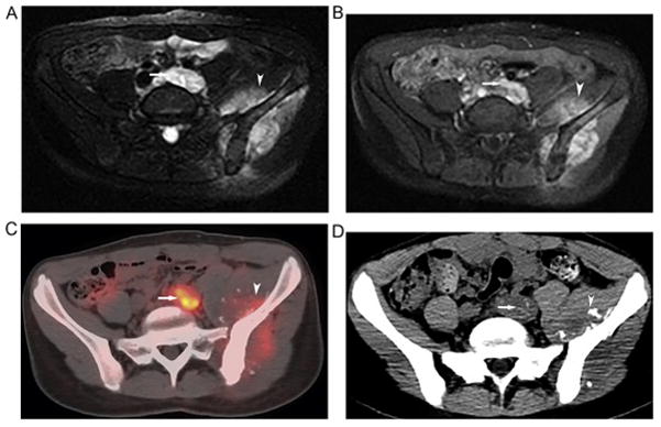
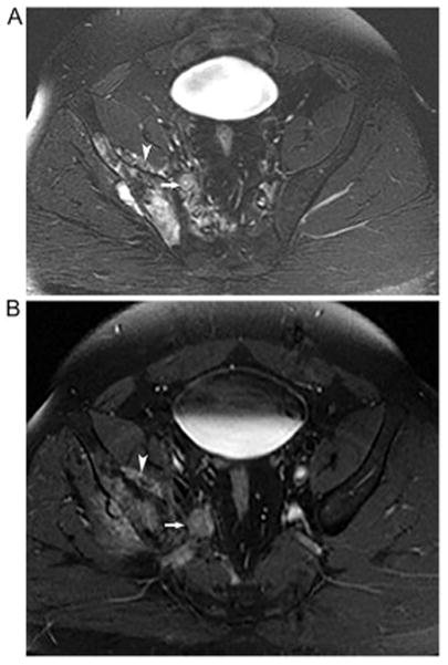
Materials and methods: Our retrospective study included all patients with primary pelvic osteosarcoma treated at our facility between January 2000 and May 2014, who were ≤ 45 years of age, and had adequate imaging studies and clinical follow up. Four radiologists evaluated for tumor in the large draining veins on initial CT, MRI and PET/CTs. A consensus evaluation by the four radiologists together with findings on operative reports, pathology reports or follow-up imaging was used as the reference standard.
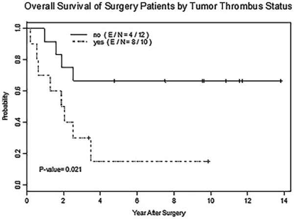
Results: Thirty-nine patients with primary pelvic osteosarcoma met final inclusion criteria. Tumor thrombus was identified in the large draining veins in 10 of the 22 (45%) patients who underwent tumor resection and 10 of the 17 (59%) who did not. In the 22 patients who underwent tumor resection, tumor thrombus was significantly associated with worse overall survival (p = 0.03).
Conclusions: Tumor thrombus in the large draining veins is identified in a significant proportion of initial imaging studies in patients with pelvic osteosarcoma, and is associated with worse overall survival in patients who undergo tumor resection.
Keywords: Large draining veins; Pelvic osteosarcoma; Tumor thrombus.
Copyright © 2018 Elsevier B.V. All rights reserved.
Figures
References
-
- Benezech S, Chabaud S, Chambon F, Dijoud F, Chotel F, Marec-Berard P. Prognostic Value of Vascular Invasion in Pediatric Osteosarcomas. Pathol Oncol Res. 2016;22:847–852. - PubMed
-
- Hoehn W, Hermanek P. Invasion of veins in renal cell carcinoma - frequency, correlation and prognosis. Eur Urol. 1983;9:276–280. - PubMed
-
- Ikai I, Arii S, Kojiro M, Ichida T, Makuuchi M, Matsuyama Y, Nakanuma Y, Okita K, Omata M, Takayasu K, Yamaoka Y. Reevaluation of prognostic factors for survival after liver resection in patients with hepatocellular carcinoma in a Japanese nationwide survey. Cancer. 2004;101:796–802. - PubMed
-
- Krasna MJ, Flancbaum L, Cody RP, Shneibaum S, Ben Ari G. Vascular and neural invasion in colorectal carcinoma. Incidence and prognostic significance. Cancer. 1988;61:1018–1023. - PubMed
-
- Kim TU, Kim S, Lee NK, Kim HJ, Han GJ, Lee JW, Baek HJ, Jeon TY, Kim HS, Park DY. Prognostic Value of Computed Tomography-Detected Extramural Venous Invasion to Predict Disease-Free Survival in Patients With Gastric Cancer. J Comput Assist Tomogr. 2016;7:7. - PubMed
MeSH terms
Grants and funding
LinkOut - more resources
Full Text Sources
Other Literature Sources
Medical
Research Materials
Miscellaneous
