. 2018 Sep 5;26(9):2107-2118.
doi: 10.1016/j.ymthe.2018.06.024. Epub 2018 Jul 5.

Extracellular Vesicles Containing IL-4 Modulate Neuroinflammation in a Mouse Model of Multiple Sclerosis

Affiliations

Extracellular Vesicles Containing IL-4 Modulate Neuroinflammation in a Mouse Model of Multiple Sclerosis

Giacomo Casella et al. Mol Ther. .

Abstract

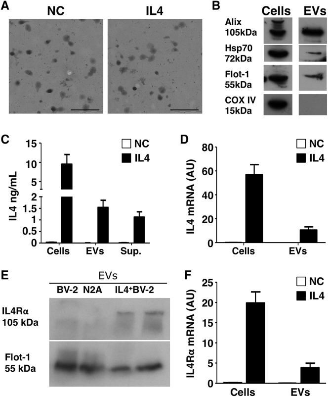
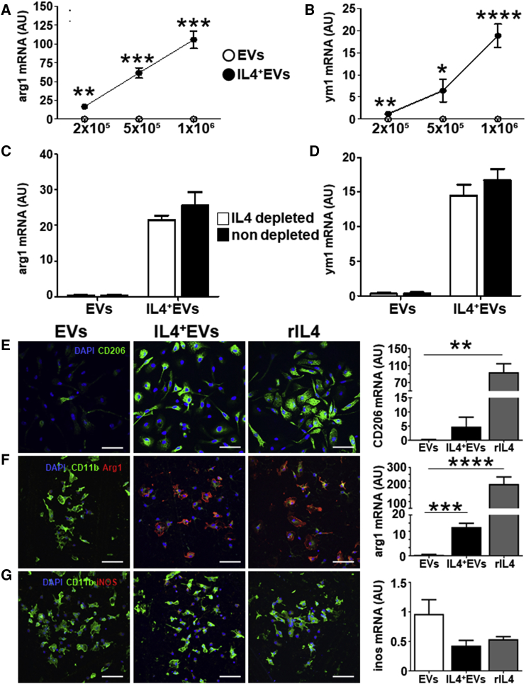
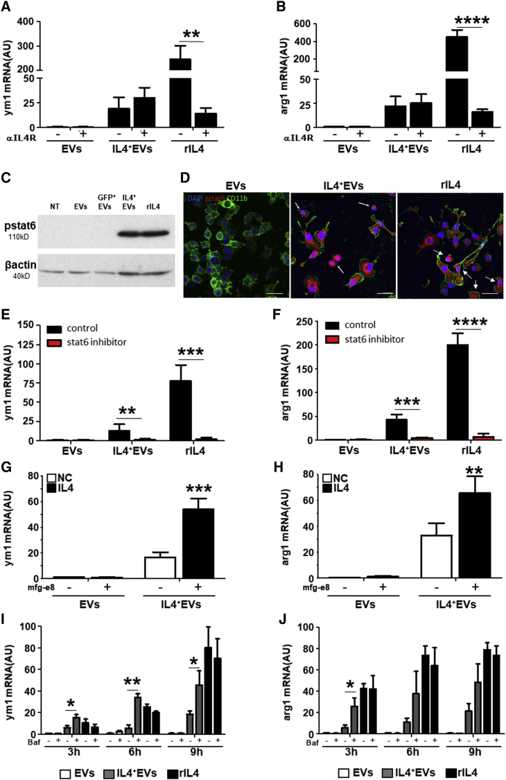
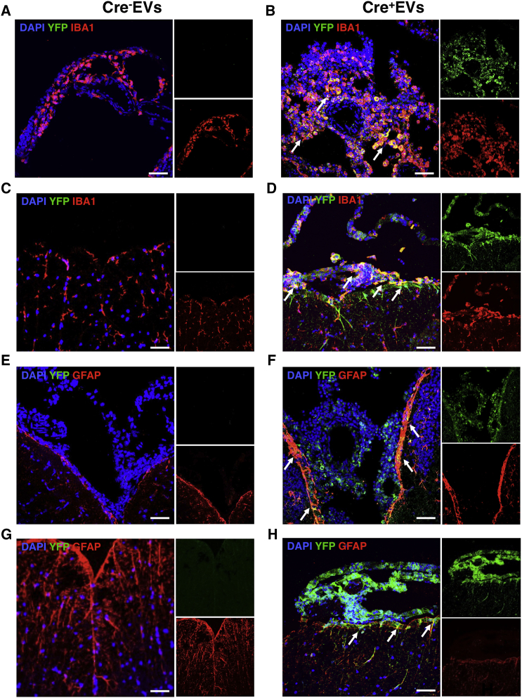
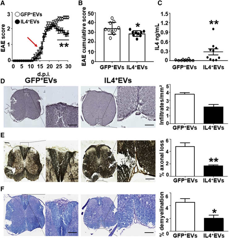
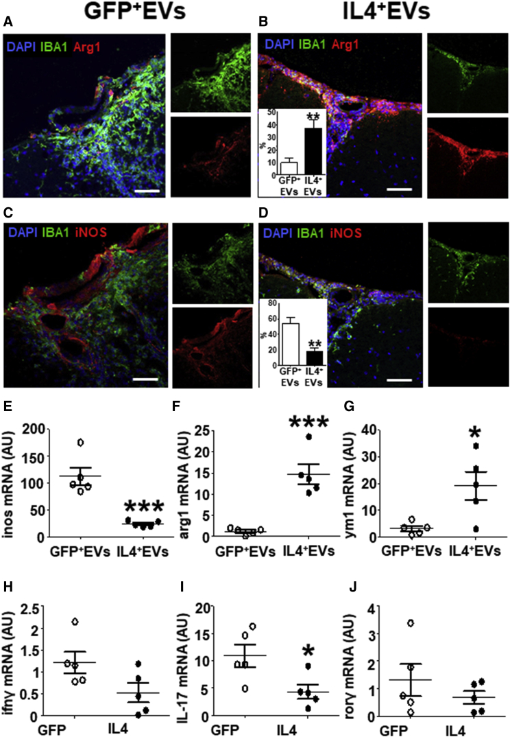
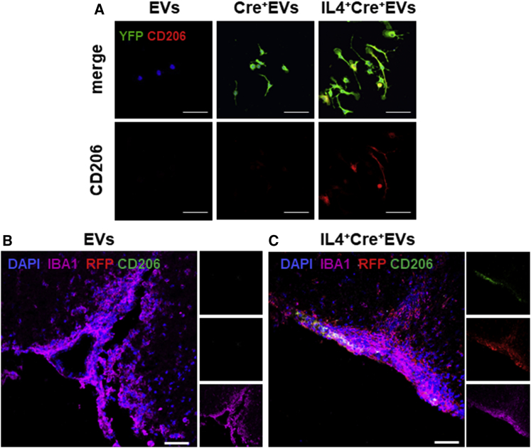
Extracellular vesicles (EVs) play a major role in cell-to-cell communication in physiological and pathological conditions, and their manipulation may represent a promising therapeutic strategy. Microglia, the parenchymal mononuclear phagocytes of the brain, modulate neighboring cells also through the release of EVs. The production of custom EVs filled with desired molecules, possibly targeted to make their uptake cell specific, and their administration in biological fluids may represent a valid approach for drug delivery. We engineered a murine microglia cell line, BV-2, to release EVs overexpressing the endogenous "eat me" signal Lactadherin (Mfg-e8) on the surface to target phagocytes and containing the anti-inflammatory cytokine IL-4. A single injection of 107 IL-4+Mfg-e8+ EVs into the cisterna magna modulated established neuroinflammation and significantly reduced clinical signs in the mouse model of multiple sclerosis, experimental autoimmune encephalomyelitis (EAE). Injected IL-4+Mfg-e8+ EVs target mainly phagocytes (i.e., macrophages and microglia) surrounding liquoral spaces, and their cargo promote the upregulation of anti-inflammatory markers chitinase 3-like 3 (ym1) and arginase-1 (arg1), significantly reducing tissue damage. Engineered EVs may represent a biological drug delivery tool able to deliver multiple functional molecules simultaneously to treat neuroinflammatory diseases.

Keywords: extracellular vesicles; neuroinflammation; phagocytes.


Figures

Figure 1
Figure 1
IL-4+ EV Characterization (A) TEM analysis of a representative EV pellet derived from IL-4+BV-2 cells (IL-4) and untransfected BV-2 cells as negative control (NC) (n = 3). Scale bars, 500 nm. (B) Western blot analysis of EVs from IL-4+BV-2 cells for EV markers (Alix, Hsp70, Flotillin-1, and COX-IV) (n = 3). (C) Measurement, by ELISA, of IL-4 in IL-4+BV-2 cell lysates (cells), IL-4+ EVs (EVs), and IL-4+BV-2 supernatants (sup.). Untransfected BV-2 cells were used as a negative control (NC) (n = 5). Bars are mean values ± SD. (D) IL-4 mRNA detected by RT-PCR in IL-4+BV-2 cells and IL-4+ EVs (IL-4) as compared to untransfected BV-2 cells (NC) (n = 5). Values are expressed as a.u. and bars represent mean values ± SD. (E) WB for IL-4Rα on EVs from untransfected BV-2 cells (BV-2), from the N2A neuronal cell line (N2A), and from IL-4+ BV-2 cells (IL-4+ BV-2) (n = 3). Protein load was controlled using flotilin-1 (Flot-1). (F) RT-PCR for IL-4Rα mRNA in EVs from untransfected BV-2 cells (NC) and IL-4+BV-2 cells (IL-4), and corresponding parental cells (n = 4). Values are expressed as a.u. and bars represent mean values ± SD. Blots in (B) and (E) have been cropped. Full pictures are presented in Figure S7.
Figure 2
Figure 2
IL-4+ EVs Modulate Recipient Cells In Vitro (A and B) RT-PCR analysis for arginase-1 (arg1) (A) and ym1 (B) in recipient BV-2 cells, treated, as indicated on the x axis, with 2 × 105, 5 × 105, and 1 × 106 IL-4+ EVs (IL-4+ EVs, closed dots) or IL-4 EVs (EVs, open dots), for 8 hr (n = 4). Values are expressed as a.u. and data points represent mean values ± SD. (C and D) To exclude IL-4 carryover, 1 × 106 IL-4+ EVs depleted of extra-vesicular IL-4 by treatment with anti-IL-4 antibody and protein G precipitation (open bars), or non-depleted 1 × 106 IL-4+ EVs (closed bars), were added to recipient BV-2 (n = 4), arg1 (C), and ym1 (D). Values are expressed as a.u. and bars represent mean values ± SD and were analyzed with two-tailed unpaired t test, *p < 0.005, **p < 0.001, ***p < 0.0001, and ****p < 0.00001. (E–G) IL-4+ EVs’ effect on primary microglia phenotype. Immunofluorescence is shown for CD206 (green, E; Arg1, red, F; iNOS, red, G) in primary microglia treated with IL-4 EVs, IL-4+ EVs (EV:cell ratio 3.5:1), and rIL-4 (20 ng/mL) for 12 hr (n = 5). Along with Arg1 and iNOS, CD11B (green) was used to identify myeloid cells and DAPI (blue) for nuclei. Levels of the corresponding mRNA (CD206, arg1, and ym1), measured by RT-PCR in the same cultures, are shown in the bar graphs on the right (n = 5). Gapdh was used as a housekeeping gene. Ym1, arg1, and inos mRNA levels are expressed as a.u.; data are expressed as means ± SD and were analyzed with the one-way ANOVA test (**p < 0.001, ***p < 0.0001, and ****p < 0.00001, E–G).
Figure 3
Figure 3
IL-4+ EV Signaling Occurs Inside the Cell and Is Dependent on STAT6 and Lysosome Efficiency (A and B) RT-PCR analysis for ym1 (A) and arginase-1 (arg1) (B) in recipient BV-2 cells treated with 1 × 106 IL-4 EVs (EVs), IL-4+ EVs (IL-4+ EVs), or recombinant IL-4 (20 ng/mL, rIL-4) for 8 hr. IL-4Rα-neutralizing antibody (1 μg/mL) was added 1 hr before treatment (n = 4). Gapdh was used as a housekeeping gene. ym1 and arg1 mRNA levels are expressed as a.u.; data are expressed as mean ± SD and were analyzed with two-tailed unpaired t test (**p < 0.001 and ****p < 0.00001). (C) Western blot for phosphorylated STAT6 (pstat6) in untreated BV-2 cells (NT) or BV-2 cells receiving 1 × 106 IL-4 EVs (EVs), GFP+ EVs, IL-4+ EVs, or 20 ng/mL recombinant IL-4 (rIL-4) for 4 hr (n = 3). Predicted molecular weight (MW) of pSTAT6 is 110 kDA. Protein load was controlled using β-actin (MW = 40 kDa). (D) Immunofluorescence for phosphorylated pstat6 (red, pstat6), CD11b (green), and nuclei (DAPI) in BV-2 cells receiving 1 × 106 IL-4 EVs (EVs), IL-4+ EVs, or rIL-4 (20 ng/mL) for 4 hr (n = 4). (E and F) The stat6 inhibitor AS1517499 (10 μM; red bars) or control medium (control; black bars) was added 1 hr before treatment of recipient BV-2 cells with IL-4 EVs, IL-4+ EVs, or rIL-4. The reaction was blocked after 5 hr, and RT-PCR for ym1 and arg1 was performed on recipient BV-2 cells (n = 5). ym1 (E) and arg1 (F) mRNA levels are expressed as a.u.; data are expressed as means ± SD and were analyzed with unpaired t test two-tailed (**p < 0.001, ***p < 0.0001, and ****p < 0.00001). (G and H) IL-4+Mfg-e8+ EVs are more efficient in modulating gene transcription compared to IL-4+ EVs in recipient BV-2 cells. RT-PCR is shown for ym1 (G) and arg1 (H) mRNA transcripts from BV-2 cells treated with 1 × 106 IL-4 or IL-4+ EVs expressing or not Mfg-8, for 8 hr (n = 5). Gapdh was used as a housekeeping gene. mRNA levels are expressed as a.u.; data are expressed as means ± SD and were analyzed with two-tailed unpaired t test (**p < 0.001 and ***p < 0.0001). (I and J) Bafilomycin (1 μM) was added for 1 hr before treatment of recipient BV-2 cells with 1 × 106 IL-4 EVs (white bars), IL-4+ EVs (gray bars), or rIL-4 (20 ng/mL; black bars) for 3, 6, and 9 hr, and RT-PCR for ym1 (I) and arg1 (J) was performed on recipient BV-2 cells (n = 5). ym1 and arg1 mRNA levels are expressed as a.u.; data are expressed as means ± SD and were analyzed with unpaired t test two tailed (*p < 0.01 and **p < 0.001). Blots (C) have been cropped. Full picture is presented in Figure S7.
Figure 4
Figure 4
Mfg-e8+ EV Delivery In Vivo R26-stop-EYFP reporter mice were intracisternally injected with 107 Mfg-e8+ EVs containing (Cre+ EVs; B, D, F, and H) or not Cre recombinase (Cre EVs; A, C, E, and G). Coronal brain (A, B, E, and F) and spinal cord sections (C, D, G, and H) were stained for YFP (green) (A–H), and for IBA1 (red; A–D) or GFAP (red; E–H) and DAPI (blue). Sections are a representation of third ventricle (A, B, E, and F) and thoracic region (C, D, G, and H). Magnification, 40×; scale bar, 50 μm; n = 3/group.
Figure 5
Figure 5
IL-4+ Mfg-e8+ EV Injection Modulates EAE and Protects EAE Mice from Tissue Damage (A) EAE mice were intracisternally injected, at clinical onset (red arrow), with 107 Mfg-8+ EVs containing GFP (GFP+ EVs, open dots) or IL-4 (IL-4+ EVs, closed dots). Dots represent mean daily EAE score; data are expressed as means ± SEM and were analyzed with two-way ANOVA Bonferroni test (**p < 0.005). n = 10/group. (B) EAE cumulative score of the same mice intracisternally injected with 107 EVs containing GFP (GFP+ EVs, open dots) or IL-4 (IL-4+ EVs, closed dots) (n = 10). (C) In the same mice, IL-4 protein was measured in the CSF by ELISA. Data are expressed as means ± SEM and were analyzed with two-tailed unpaired t test (**p < 0.01) (n = 10). (D–F) Histological analysis of the spinal cord of EAE mice injected with 107 GFP+Mfg-e8+ (GFP+ EVs) or IL-4+Mfg-e8+ EVs (IL-4+ EVs) (n = 5). Adjacent sections are stained to detect inflammatory infiltrates (H&E, D), axonal loss (Bielshowsky, E), and demyelination (Kluver Barrera, F). Magnification, 20×; n = 5/group; scale bar, 100 μm. Column bars represent mean values ± SD; *p < 0.05 and **p < 0.001 (two-tailed unpaired t test).
Figure 6
Figure 6
IL-4+Mfg-e8+ EV Injection in EAE Mice Induces Anti-inflammatory Markers on CNS Phagocytes (A–D) Immunofluorescence for Arg1 (red; A and B) and iNOS (red; C and D) in spinal cord sections of EAE mice injected with 107 GFP+ (GFP+ EVs) or IL-4+ Mfg-e8+ EVs (IL-4+ EVs) (n = 5). IBA1 (green) was used to identify myeloid cells and DAPI (blue) for nuclei. Percentage of double-positive IBA1+Arg1+ and IBA1+iNOS+ cells on total IBA1+ cells are shown in the insets of (B)–(D) in IL-4-treated (closed bars) or GFP-treated (open bars) mice. Data are expressed as means ± SD and were analyzed with two-tailed unpaired t test (**p < 0.01). (E–J) RT-PCR for inos (E), arg1 (F), and ym1 mRNA (G) was performed on CNS-infiltrating CD11b+ cells, whereas RT-PCR for ifnγ (H), il-17 (I), and rorγ mRNAs (J) was performed on CNS-infiltrating CD4+cells from EAE mice intracisternally injected with 107 GFP+ (GFP+ EVs) or IL-4+ Mfg-e8+ EVs (IL-4+ EVs) (n = 5). Gapdh was used as a housekeeping gene. Data are shown as a.u. and were analyzed with two-tailed unpaired t test (*p < 0.05 and ***p < 0.001).
Figure 7
Figure 7
IL-4+Mfg-e8+ EVs Induce Anti-inflammatory Markers Only in Recipient Cells (A) Immunofluorescence for YFP (green), CD206 (red), and DAPI (blue) on bone marrow macrophages from R26-stop-EYFP mice exposed for 24 hr to EVs, Cre+ EVs, and IL-4+Cre+ EVs (n = 4). 60×; scale bar, 20 μm. (B and C) In vivo, IL-4+Mfg-e8+Cre+ EVs induce the upregulation of the anti-inflammatory marker CD206 only in IBA1+ cells that had recombined the reporter locus (C), compared to control EAE mice treated with IL-4Mfg-e8+Cre+ EVs (B); sections were stained for IBA1 (magenta), RFP (red), CD206 (green), and DAPI (blue). Magnification, 40×; scale bar, 50 μm; n = 3/group.

References

    1. Théry C., Ostrowski M., Segura E. Membrane vesicles as conveyors of immune responses. Nat. Rev. Immunol. 2009;9:581–593. - PubMed
    1. Porro C., Trotta T., Panaro M.A. Microvesicles in the brain: Biomarker, messenger or mediator? J. Neuroimmunol. 2015;288:70–78. - PubMed
    1. Cocucci E., Racchetti G., Meldolesi J. Shedding microvesicles: artefacts no more. Trends Cell Biol. 2009;19:43–51. - PubMed
    1. Mulcahy L.A., Pink R.C., Carter D.R. Routes and mechanisms of extracellular vesicle uptake. J. Extracell. Vesicles. 2014;3 - PMC - PubMed
    1. Lässer C., Théry C., Buzás E.I., Mathivanan S., Zhao W., Gho Y.S., Lötvall J. The International Society for Extracellular Vesicles launches the first massive open online course on extracellular vesicles. J. Extracell. Vesicles. 2016;5:34299. - PMC - PubMed

Publication types

MeSH terms