. 2018 Sep 3;150(9):1317-1331.
doi: 10.1085/jgp.201811993. Epub 2018 Jul 17.

Propofol inhibits the voltage-gated sodium channel NaChBac at multiple sites

Affiliations

Propofol inhibits the voltage-gated sodium channel NaChBac at multiple sites

Yali Wang et al. J Gen Physiol. .

Abstract

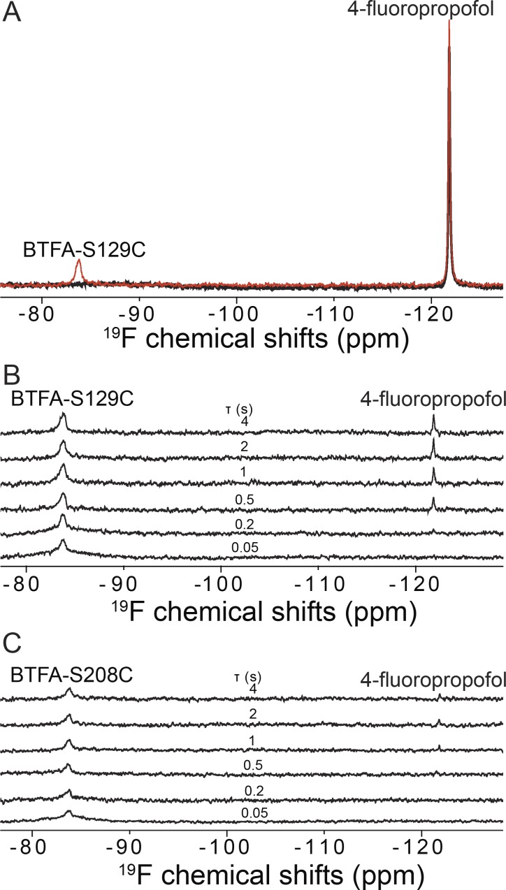
Voltage-gated sodium (NaV) channels are important targets of general anesthetics, including the intravenous anesthetic propofol. Electrophysiology studies on the prokaryotic NaV channel NaChBac have demonstrated that propofol promotes channel activation and accelerates activation-coupled inactivation, but the molecular mechanisms of these effects are unclear. Here, guided by computational docking and molecular dynamics simulations, we predict several propofol-binding sites in NaChBac. We then strategically place small fluorinated probes at these putative binding sites and experimentally quantify the interaction strengths with a fluorinated propofol analogue, 4-fluoropropofol. In vitro and in vivo measurements show that 4-fluoropropofol and propofol have similar effects on NaChBac function and nearly identical anesthetizing effects on tadpole mobility. Using quantitative analysis by 19F-NMR saturation transfer difference spectroscopy, we reveal strong intermolecular cross-relaxation rate constants between 4-fluoropropofol and four different regions of NaChBac, including the activation gate and selectivity filter in the pore, the voltage sensing domain, and the S4-S5 linker. Unlike volatile anesthetics, 4-fluoropropofol does not bind to the extracellular interface of the pore domain. Collectively, our results show that propofol inhibits NaChBac at multiple sites, likely with distinct modes of action. This study provides a molecular basis for understanding the net inhibitory action of propofol on NaV channels.


Figures

Figure 1.
Figure 1.
In vivo anesthetizing concentrations of propofol and 4-fluoropropofol in albino X. laevis tadpoles are identical. The percentage of immobilized tadpoles is plotted as a function of propofol and 4-fluoropropofol concentrations. The solid lines are best fit to the data by using Eq. 4. Error bars show the SEMs from three independent measurements with 10 tadpoles per measurement at each drug concentration.
Figure 2.
Figure 2.
4-Fluoropropofol modulates voltage-dependent activation and inactivation of NaChBac. (A) Representative paired current families in the absence (control) and presence of 4 µM 4-fluoropropofol. (B) Paired currents at +20 mV. The 4-µM 4-fluoropropofol trace is shown scaled to its respective control Ipeak. The 40-µM 4-fluoropropofol trace is shown both as a raw current and scaled to its respective control Ipeak. (C) Time constants of inactivation (τ) versus voltage of control (n = 20) and 4-fluoropropofol at 4 and 40 µM (n = 8–12). 4-Fluoropropofol reduced τInactivation at both concentrations and all voltages (P < 0.0001, paired t test). (D and E) Normalized G-V (D) and prepulse (E) inactivation curves of control and 4-fluoropropofol at the indicated concentrations (n = 5–12). Corresponding midpoints (V1/2) of activation and inactivation are shown below the corresponding curves. Means are indicated in magenta. Data are reported as mean ± SEM from n independent measurements.
Figure 3.
Figure 3.
In silico predicted propofol binding sites in NaChBac. (A) Top and side views of a NaChBac structure model with putative propofol binding sites identified by molecular docking (cyan spheres, occupancy ≥ 10%). (B and C) Zoomed-in views of the lowest energy conformation from docking calculations of propofol (cyan) bound to NaChBac inside the pore at the selectivity filter (B) and above the activation gate (C). Residues in close proximity to the docked propofol are shown as sticks labeled with the corresponding color. Note that residues selected for 19F labeling in STD NMR experiments (T189, I223, F227) are within the binding sites but unlikely to be essential for propofol binding to these regions. (D) Top and side views of NaChBac from the final frame of the 1-µs flooding MD simulation with putative propofol-binding sites (cyan spheres, occupancy ≥ 90%). (E and F) Zoomed-in views of propofol bound to NaChBac in the final frame of the 1-µs flooding MD simulation in the apex of the voltage-sensing domain (E) and an intersubunit site at the intracellular interface (F). Residues in close proximity to the docked propofol are shown as sticks labeled with the corresponding color. Note that residues selected for 19F labeling in STD NMR experiments (N36, V40, S129, L150) are within the binding sites but unlikely to be essential for propofol binding to these regions.
Figure 4.
Figure 4.
Effects of single-cysteine mutations on NaChBac modulation by 4-fluoropropofol. (A) Paired G-V and prepulse inactivation curves without (black) and with (light blue) 4 µM 4-fluoropropofol (n = 4–13) for WT and each NaChBac mutant. (B and C) ΔV1/2 values of activation (B) and inactivation (C) induced by 4 µM 4-fluoropropofol for WT and NaChBac mutants. The ΔV1/2 values of activation were not significantly different between WT and the mutants. Of the four mutants, only S129C shows significantly different ΔV1/2 of inactivation from the WT (P = 0.0384, one-way ANOVA with Bonferroni post hoc correction). (D) Fold-change in time constants (τ) of inactivation (τ4-fluoropropofolcontrol) at +20 mV induced by 4 µM 4-fluoropropofol. Time constants were derived from the decaying phase of the Na+ currents, which were well described by the single exponential function. For all mutants, the fold-change in τInactivation caused by 4 µM 4-fluoropropofol was not significantly different from that of WT (one-way ANOVA with Bonferroni post hoc correction). Data are reported as mean ± SEM from n independent measurements.
Figure 5.
Figure 5.
19F-NMR measurements of 4-fluoropropofol binding to NaChBac. (A) Representative 19F-NMR spectra of the BTFA-labeled S129C NaChBac mutant in the presence of 200 µM 4-fluoropropofol with selective on- (black, Ion) and off-resonance (red, Ioff) saturation of the BTFA peak. The saturation time was 2 s. (B) Stack plot of 19F STD NMR spectra showing that 4-fluoropropofol interacts specifically with the BTFA labeled at S129C for intermolecular saturation transfer to be built up at longer saturation time (τ). (C) Stack plot of 19F STD NMR spectra showing that 4-fluoropropofol has no measurable interaction with the negative control BTFA-labeled S208C NaChBac.
Figure 6.
Figure 6.
Quantitative analysis of 19F NMR STD build-up from individual mutation sites in NaChBac to 4-fluoropropofol. Mutations are grouped by four regions: (A) the extracellular interface (S208), (B) the S4–S5 linker region (L150C, S129C), (C) the apex of the voltage sensing domain (N36C, V40C), and (D) the pore region (F227C, I223, T189C). The solid lines are the best fit to the data by using the two-parameter equation (Eq. 1), yielding the cross-relaxation rate constant and the saturating magnetization transfer from the 19F labels on NaChBac to 4-fluoropropofol. Error bars are uncertainties calculated from the root-mean-squared noise-to-signal ratios in the on- and off-resonance 19F NMR spectra.
Figure 7.
Figure 7.
Local anesthetic inhibition of 4-fluoropropofol 19F NMR STD signal in the pore of NaChBac. (A and B) Stack plots of 19F STD NMR spectra for 100 µM 4-fluoropropofol interacting with the BTFA labeled at I223C in the absence (A) and presence (B) of 50 µM etidocaine, a local anesthetic. (C) 19F NMR STD accumulation in the absence (black) and presence (red) of 50 µM etidocaine. The solid lines are the best fit to the data by using the two-parameter equation (Eq. 1), yielding the cross-relaxation rate constant and the saturating magnetization transfer from the 19F labels on I223C in NaChBac to 4-fluoropropofol. Error bars are uncertainties calculated from the root-mean-square noise-to-signal ratios in the on- and off-resonance 19F NMR spectra.

Comment in

  • Propofol's paradox, explained.
    Sedwick C. Sedwick C. J Gen Physiol. 2018 Sep 3;150(9):1231-1232. doi: 10.1085/jgp.201812197. Epub 2018 Aug 16. J Gen Physiol. 2018. PMID: 30115662 Free PMC article.

References

    1. Amarouch M.Y., and Abriel H.. 2015. Cellular hyper-excitability caused by mutations that alter the activation process of voltage-gated sodium channels. Front. Physiol. 6:45 10.3389/fphys.2015.00045 - DOI - PMC - PubMed
    1. Angulo J., Enríquez-Navas P.M., and Nieto P.M.. 2010. Ligand-receptor binding affinities from saturation transfer difference (STD) NMR spectroscopy: the binding isotherm of STD initial growth rates. Chemistry. 16:7803–7812. 10.1002/chem.200903528 - DOI - PubMed
    1. Barber A.F., Carnevale V., Raju S.G., Amaral C., Treptow W., and Klein M.L.. 2012. Hinge-bending motions in the pore domain of a bacterial voltage-gated sodium channel. Biochim. Biophys. Acta. 1818:2120–2125. 10.1016/j.bbamem.2012.05.002 - DOI - PMC - PubMed
    1. Barber A.F., Carnevale V., Klein M.L., Eckenhoff R.G., and Covarrubias M.. 2014. Modulation of a voltage-gated Na+ channel by sevoflurane involves multiple sites and distinct mechanisms. Proc. Natl. Acad. Sci. USA. 111:6726–6731. 10.1073/pnas.1405768111 - DOI - PMC - PubMed
    1. Boiteux C., Vorobyov I., French R.J., French C., Yarov-Yarovoy V., and Allen T.W.. 2014. Local anesthetic and antiepileptic drug access and binding to a bacterial voltage-gated sodium channel. Proc. Natl. Acad. Sci. USA. 111:13057–13062. 10.1073/pnas.1408710111 - DOI - PMC - PubMed

Publication types

LinkOut - more resources