. 2018 Jul 17;11(539):eaao3964.
doi: 10.1126/scisignal.aao3964.

Unique BIR domain sets determine inhibitor of apoptosis protein-driven cell death and NOD2 complex signal specificity

Affiliations

Unique BIR domain sets determine inhibitor of apoptosis protein-driven cell death and NOD2 complex signal specificity

Steven M Chirieleison et al. Sci Signal. .

Abstract

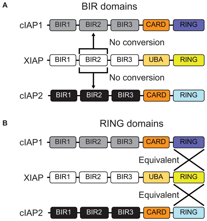
The mammalian IAPs, X-linked inhibitor of apoptosis protein (XIAP) and cellular inhibitor of apoptosis protein 1 and 2 (cIAP1 and cIAP2), play pivotal roles in innate immune signaling and inflammatory homeostasis, often working in parallel or in conjunction at a signaling complex. IAPs direct both nucleotide-binding oligomerization domain-containing 2 (NOD2) signaling complexes and cell death mechanisms to appropriately regulate inflammation. Although it is known that XIAP is critical for NOD2 signaling and that the loss of cIAP1 and cIAP2 blunts NOD2 activity, it is unclear whether these three highly related proteins can compensate for one another in NOD2 signaling or in mechanisms governing apoptosis or necroptosis. This potential redundancy is critically important, given that genetic loss of XIAP causes both very early onset inflammatory bowel disease and X-linked lymphoproliferative syndrome 2 (XLP-2) and that the overexpression of cIAP1 and cIAP2 is linked to both carcinogenesis and chemotherapeutic resistance. Given the therapeutic interest in IAP inhibition and the potential toxicities associated with disruption of inflammatory homeostasis, we used synthetic biology techniques to examine the functional redundancies of key domains in the IAPs. From this analysis, we defined the features of the IAPs that enable them to function at overlapping signaling complexes but remain independent and functionally exclusive in their roles as E3 ubiquitin ligases in innate immune and inflammatory signaling.


Figures

Fig. 1
Fig. 1. Attempts to generate triple IAP-knockout cells are unsuccessful and result in up-regulation of the remaining IAP protein
HT29 cells were stably transduced with CRISPR/Cas9 constructs targeting either cIAP1, XIAP, or cIAP2 to generate single-, double-, or triple-knockout cell lines as indicated. Single and double IAP knockouts were readily detected (top and middle), but no triple knockouts were detected (“?”, bottom). Neg, nontargeted CRISPR/Cas9 control; Neg*, the same CRISPR/Cas9 cell line was used as a Western blot control in the right double-knockout blot and the triple-knockout blot. Single-knockout: cIAP1, n = 12 clones; XIAP, n = 11 clones; and cIAP2, n = 13 clones. Double-knockout: XIAP/cIAP1, n = 13 clones and XIAP/cIAP2, n = 13 clones. Triple knockout, n = clones.
Fig. 2
Fig. 2. cIAP1 and cIAP2 cannot fully compensate for XIAP loss
(A) Schematic of XIAP, cIAP1, and cIAP2 proteins reconstituted to XIAP-knockout cell lines. (B) Relative NOD2-activated NF-κB–driven luciferase activity in HEK293T cells reconstituted with the indicated IAP protein. Relative activity normalized to an XIAP activity of 100. *P ≤ 0.05 compared to empty by one-way analysis of variance (ANOVA) with Bonferroni’s multiple comparisons test. Data are mean ± SEM from n = 6 experimental replicates performed on different days. (C) IRG1 expression assayed by real-time quantitative polymerase chain reaction (qPCR) in DC2.4 XIAP–knockout cells reconstituted with empty vector or the indicated IAP protein after stimulation with L18-MDP (10 μg/ml) for 3 hours. *P ≤ 0.05 compared to empty 3-hour time point by one-way ANOVA with Bonferroni’s multiple comparisons test. Data are mean ± SEM from n = 3 experimental replicates performed on different days. (D) iBMDM XIAP–knockout cell lines re-constituted with the indicated IAP proteins were stimulated with L18-MDP (10 μg/ml) for 0, 30, 60, and 120 min. Cellular lysates were examined by Western blot for NF-κB signaling using the indicated antibodies. Lysates from XIAP at the 0- and 120-min time points of stimulation are included on all blots for comparison. Representative blots from one of three separate experiments are shown. (E) XIAP-knockout iBMDM cells reconstituted with the indicated IAP protein were treated with TNF (10 ng/ml) or TNF (10 ng/ml) and GDC-0152 (200 nM) for 24 hours, and cell viability was quantified by methylene blue staining. *P ≤ 0.05 compared to empty TNF + GDC-0152 by one-way ANOVA with Bonferroni’s multiple comparisons test. Data are mean ± SEM from n = 3 experimental replicates performed on different days. (F) iBMDM XIAP–knockout cells reconstituted with the indicated IAP protein were treated with TNF (10 ng/ml) alone or in combination with GDC-0152 (200 nM) as indicated for 8 hours, and cellular lysates were assayed by Western blot for caspase-3 cleavage. Representative blots from one of three separate experiments are shown. GAPDH, glyceraldehyde-3-phosphate dehydrogenase; p-IκBα, phosphorylated IκBα.
Fig. 3
Fig. 3. cIAP1 carrying XIAP’s BIR2 domain can partially compensate for XIAP loss
(A) Schematic of XIAP and cIAP1 and cIAP2 chimeric proteins in which the BIR2 of the native cIAP was replaced with the BIR2 domain of XIAP (X-BIR2). (B) HEK293T cells reconstituted with XIAP or a cIAP1/2 chimeric protein were assayed for relative NF-κB luciferase activity in response to NOD2 overexpression. Relative units were normalized to an XIAP activity of 100. *P ≤ 0.05 compared to empty by one-way ANOVA with Bonferroni’s multiple comparisons test. Data are mean ± SEM from n = 6 experimental replicates performed on different days. (C) DC2.4 XIAP–knockout cell line reconstituted with the indicated IAP variant was assayed for IRG1 expression by real-time qPCR after stimulation with L18-MDP (10 μg/ml) for 3 hours. *P ≤ 0.05 compared to empty 3-hour time point by one-way ANOVA with Bonferroni’s multiple comparisons test. Data are mean ± SEM from n = 3 experimental replicates performed on different days. (D) After L18-MDP (10 μg/ml) treatment for 0, 30, 60, or 120 min, cellular lysates from iBMDM cells reconstituted with the indicated IAP variants were examined for NF-κB signaling by Western blot using the indicated antibodies. Lysates from XIAP at the 0- and 120-min time points of stimulation are included on all blots for comparison. Representative blots from one of three separate experiments are shown. (E) XIAP or cIAP1/2 chimeric proteins expressed in XIAP-knockout iBMDM cells were assayed for their ability to suppress cell death after TNF (10 ng/ml) or TNF and GDC-0152 (200 nM) stimulation. After 24 hours of treatment, cell viability was assayed by methylene blue staining. *P ≤ 0.05 compared to empty TNF + GDC-0152 by one-way ANOVA with Bonferroni’s multiple comparisons test. Data are mean ± SEM from n = 3 experimental replicates performed on different days. (F) Cell lysates from iBMDM cells stimulated with TNF (10 ng/ml) and GDC-0152 (200 nM) as indicated for 8 hours were assayed by Western blot for caspase-3 cleavage. Representative blots from one of three separate experiments are shown.
Fig. 4
Fig. 4. The IAP RING domains of XIAP, cIAP1, and cIAP2 are functionally redundant
(A) Schematic of XIAP and XIAP chimeric proteins containing the RING domain from either cIAP1 or cIAP2. (B) Relative NF-κB–driven luciferase activity after NOD2 overexpression in HEK293T cells reconstituted with the indicated XIAP protein variant. Relative units were normalized to an XIAP activity of 100. *P ≤ 0.05 compared to empty by one-way ANOVA with Bonferroni’s multiple comparisons test. Data are mean ± SEM from n = 6 experimental replicates performed on different days. (C) DC2.4 XIAP–knockout cells reconstituted with empty vector or the indicated XIAP protein stimulated with L18-MDP (10 μg/ml) for 3 hours and assayed for the expression of IRG1 by real-time qPCR. *P ≤ 0.05 compared to empty 3-hour time point by one-way ANOVA with Bonferroni’s multiple comparisons test. Data are mean ± SEM from n = 3 experimental replicates performed on different days. (D) iBMDM XIAP–knockout cell lines reconstituted with the indicated XIAP variants were stimulated with L18-MDP (10 μg/ml) for 0, 30, 60, 90, and 120 min, and cellular lysates were examined for NF-κB signaling with the indicated antibodies. Representative blots from one of three separate experiments are shown. (E) iBMDM cells reconstituted with the indicated XIAP variants were stimulated with TNF (10 ng/ml) or TNF and GDC-0152 (200 nM) for 24 hours, and cell viability was quantified by methylene blue staining. *P ≤ 0.05 compared to empty TNF + GDC-0152 by one-way ANOVA with Bonferroni’s multiple comparisons test. Data are mean ± SEM from n = 3 experimental replicates performed on different days. (F) After stimulation with TNF (10 ng/ml) and GDC-0152 (200 nM) as indicated for 8 hours, cellular lysates were examined for induction of apoptosis by Western blot for caspase-3. Representative blots from one of three separate experiments are shown.
Fig. 5
Fig. 5. Schematic demonstrating the unique and redundant functional domains of the IAP proteins
(A) Swapping XIAP’s BIR2 domain into cIAP1 or cIAP2 results in no conversion of protein function. (B) Interchange of the RING domains of cIAP1 and cIAP2 with XIAP results in proteins with equivalent function.

References

    1. Crook NE, Clem RJ, Miller LK. An apoptosis-inhibiting baculovirus gene with a zinc finger-like motif. J Virol. 1993;67:2168–2174. - PMC - PubMed
    1. Uren AG, Coulson EJ, Vaux DL. Conservation of baculovirus inhibitor of apoptosis repeat proteins (BIRPs) in viruses, nematodes, vertebrates and yeasts. Trends Biochem Sci. 1998;23:159–162. - PubMed
    1. Duckett CS, Nava VE, Gedrich RW, Clem RJ, Van Dongen JL, Gilfillan MC, Shiels H, Hardwick JM, Thompson CB. A conserved family of cellular genes related to the baculovirus iap gene and encoding apoptosis inhibitors. EMBO J. 1996;15:2685–2694. - PMC - PubMed
    1. Deveraux QL, Takahashi R, Salvesen GS, Reed JC. X-linked IAP is a direct inhibitor of cell-death proteases. Nature. 1997;388:300–304. - PubMed
    1. Salvesen GS, Duckett CS. IAP proteins: Blocking the road to death’s door. Nat Rev Mol Cell Biol. 2002;3:401–410. - PubMed

Publication types

Substances