. 2018 Sep 14;293(37):14192-14199.
doi: 10.1074/jbc.RA118.003990. Epub 2018 Jul 17.

Plasminogen activation triggers transthyretin amyloidogenesis in vitro

Affiliations

Plasminogen activation triggers transthyretin amyloidogenesis in vitro

P Patrizia Mangione et al. J Biol Chem. .

Abstract

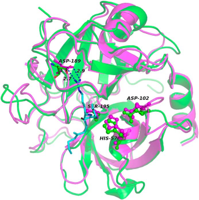
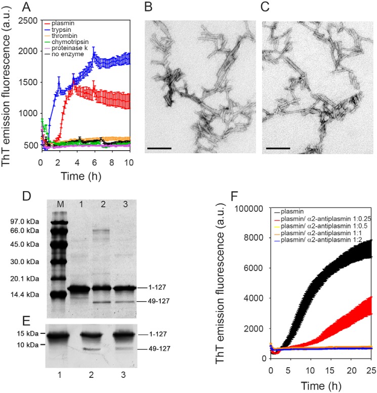
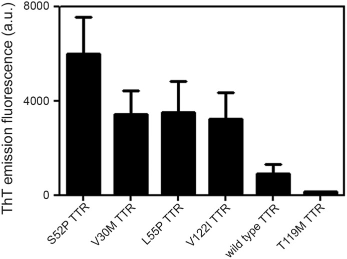
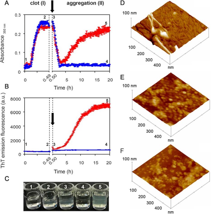
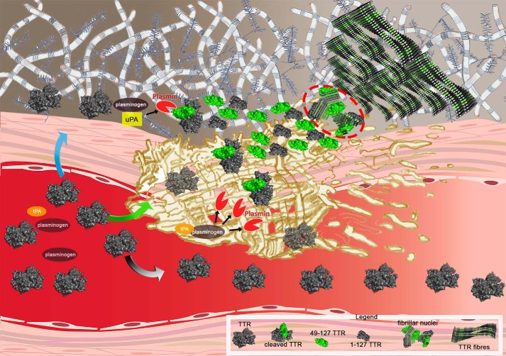
Systemic amyloidosis is a usually fatal disease caused by extracellular accumulation of abnormal protein fibers, amyloid fibrils, derived by misfolding and aggregation of soluble globular plasma protein precursors. Both WT and genetic variants of the normal plasma protein transthyretin (TTR) form amyloid, but neither the misfolding leading to fibrillogenesis nor the anatomical localization of TTR amyloid deposition are understood. We have previously shown that, under physiological conditions, trypsin cleaves human TTR in a mechano-enzymatic mechanism that generates abundant amyloid fibrils in vitro In sharp contrast, the widely used in vitro model of denaturation and aggregation of TTR by prolonged exposure to pH 4.0 yields almost no clearly defined amyloid fibrils. However, the exclusive duodenal location of trypsin means that this enzyme cannot contribute to systemic extracellular TTR amyloid deposition in vivo Here, we therefore conducted a bioinformatics search for systemically active tryptic proteases with appropriate tissue distribution, which unexpectedly identified plasmin as the leading candidate. We confirmed that plasmin, just as trypsin, selectively cleaves human TTR between residues 48 and 49 under physiological conditions in vitro Truncated and full-length protomers are then released from the native homotetramer and rapidly aggregate into abundant fibrils indistinguishable from ex vivo TTR amyloid. Our findings suggest that physiological fibrinolysis is likely to play a critical role in TTR amyloid formation in vivo Identification of this surprising intersection between two hitherto unrelated pathways opens new avenues for elucidating the mechanisms of TTR amyloidosis, for seeking susceptibility risk factors, and for therapeutic innovation.

Keywords: amyloid; amyloid fibrillogenesis; amyloidogenesis; fibril; mechano-enzymatic mechanism; protease; protein aggregation; systemic amyloidosis; tissue plasminogen activator (tPA); transthyretin.


Conflict of interest statement

The authors declare that they have no conflicts of interest with the contents of this article

Figures

Figure 1.
Figure 1.
Structural and functional similarities between trypsin and plasmin in complex with the peptide P3-P3′ corresponding to sequence 46–51 of TTR. The backbones of trypsin (magenta; PDB ID:3D65) and plasmin (green; PDB ID: 3UIR) are overlaid; the catalytic triad, in ball and stick, with the Asp residue, in sticks, that lead to the correct orientation of the Lys-substrate (Lys-48 in TTR) are specifically highlighted. The numbering refers to trypsin residues. The P3-P3′ peptide backbone of textilinin-1 in the complex with plasmin is shown in cyan. The side chain of Lys in position P1 is also represented in sticks with the distances from Asp-189. For clarity the corresponding peptide complexed to trypsin is not shown.
Figure 2.
Figure 2.
Plasmin-mediated amyloid fibrillogenesis of S52P TTR. A, increase in ThT emission fluorescence for S52P TTR incubated in the presence of plasmin compared with trypsin. No amyloid-specific ThT signal was seen after incubation of S52P TTR with thrombin, chymotrypsin, or proteinase K. B and C, negatively stained transmission electron micrographs of S52P TTR amyloid fibrils formed in the presence of trypsin (B) or plasmin (C). Scale bar, 100 nm. D, 15% SDS-PAGE under reducing conditions. M, marker proteins (14.4, 20.1, 30.0, 45.0, 66.0, and 97.0 kDa); lane 1, S52P TTR at time 0; lane 2, S52P TTR fibrils formed in the presence of trypsin; and lane 3, S52P TTR fibrils formed in the presence of plasmin. E, immunoblot analysis of samples separated in 15% SDS-PAGE (see lanes 1, 2, and 3 in D). Position of marker proteins at 15 and 10 kDa are indicated. F, inhibition by α2-antiplasmin of fibril formation by S52P TTR mediated by 20 ng/μl plasmin. The data were normalized to the ThT signal plateau in the samples without α2-antiplasmin. Mean ± S.D. of three replicates is shown. a.u., arbitrary units.
Figure 3.
Figure 3.
Plasmin-mediated fibrillogenesis. Relative ThT emission fluorescence intensities of TTR samples at 1 mg/ml after 25 h incubation with shaking in the presence of plasmin at an enzyme:substrate ratio of 1:50. Mean ± S.D. of three replicates is shown.
Figure 4.
Figure 4.
From fibrin to fibrils. A and B, spectrophotometric absorbance/light scattering at 350 nm (A) and amyloid-specific ThT emission fluorescence (B) during clotting of fibrinogen to fibrin (phase I) followed by fibrinolysis in the presence of S52P TTR (red) or of the highly stable T119M TTR variant (blue) (phase II). Following fibrinolysis, increase in turbidity and ThT were observed in the presence of S52P TTR whereas neither of these signals increased when T119M TTR was present instead. Arrows indicate addition of tPA, plasminogen, and TTR. The results shown are the mean ± S.D. of three independent experiments. C, wells containing a solution of fibrinogen in the presence of 1) thrombin and 2) fibrin clot; 3) a solution of tPA, plasminogen, and TTR layered over the clot surface; 4) fibrinolysis with no further aggregation in the presence of T119M TTR; 5) fibrinolysis in the presence of S52P TTR showing the turbidity of amyloid fibril formation. D–F, surface plots of topographic tapping mode AFM images showing (D) the presence of fibrillar structures in the sample containing clot, tPA, plasminogen, and S52P TTR; (E and F) the presence of globular structures in samples containing clot, tPA, and plasminogen (E) in the presence of T119M TTR or (F) in the absence of any TTR isoform.
Figure 5.
Figure 5.
From fibrin to fibrils. Shown is a cartoon of the putative flow of events leading to TTR amyloid fibril formation caused by plasmin cleavage within the physiological scenario of fibrin formation and plasminogen activation. Circulating TTR can diffuse toward the extracellular compartment (blue arrow), be entrapped in the fibrin clot (green arrow), or escape from it (gray arrow). In the presence of activated plasminogen both in the presence of uPA (extracellular compartment) and tPA (within the clot), tetrameric TTR may be cleaved and then rapidly dissociate into a mixture of the truncated residue 49–127 fragment (green) and full-length protomers (gray subunit). The released subunits may generate the fibrillar nuclei (highlighted within the red circle) that then aggregate into amyloid fibrils, which accumulate in the extracellular space. The legend at the bottom of the figure identifies all the TTR species.

References

    1. Pepys M. B. (2009) A molecular correlate of clinicopathology in transthyretin amyloidosis. J. Pathol. 217, 1–3 10.1002/path.2467 - DOI - PubMed
    1. Hammarström P., Schneider F., and Kelly J. W. (2001) Trans-suppression of misfolding in an amyloid disease. Science 293, 2459–2462 10.1126/science.1062245 - DOI - PubMed
    1. Mangione P. P., Porcari R., Gillmore J. D., Pucci P., Monti M., Porcari M., Giorgetti S., Marchese L., Raimondi S., Serpell L. C., Chen W., Relini A., Marcoux J., Clatworthy I. R., Taylor G. W., et al. (2014) Proteolytic cleavage of Ser52Pro variant transthyretin triggers its amyloid fibrillogenesis. Proc. Natl. Acad. Sci. U.S.A. 111, 1539–1544 10.1073/pnas.1317488111 - DOI - PMC - PubMed
    1. Klimtchuk E. S., Prokaeva T., Frame N. M., Abdullahi H. A., Spencer B., Dasari S., Cui H., Berk J. L., Kurtin P. J., Connors L. H., and Gursky O. (2018) Unusual duplication mutation in a surface loop of human transthyretin leads to an aggressive drug-resistant amyloid disease. Proc. Natl. Acad. Sci. U.S.A. 115, E6428–E6436 10.1073/pnas.1802977115 - DOI - PMC - PubMed
    1. Marcoux J., Mangione P. P., Porcari R., Degiacomi M. T., Verona G., Taylor G. W., Giorgetti S., Raimondi S., Sanglier-Cianferani S., Benesch J. L., Cecconi C., Naqvi M. M., Gillmore J. D., Hawkins P. N., Stoppini M., Robinson C. V., Pepys M. B., and Bellotti V. (2015) A novel mechano-enzymatic cleavage mechanism underlies transthyretin amyloidogenesis. EMBO Mol. Med. 7, 1337–1349 10.15252/emmm.201505357 - DOI - PMC - PubMed

Publication types