Review
. 2018 Jul 17;7(7):79.
doi: 10.3390/cells7070079.

Chlamydomonas Basal Bodies as Flagella Organizing Centers

Affiliations
Review

Chlamydomonas Basal Bodies as Flagella Organizing Centers

Jenna Lynne Wingfield et al. Cells. .

Abstract

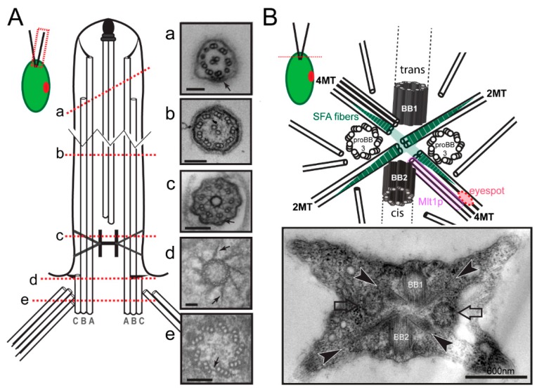
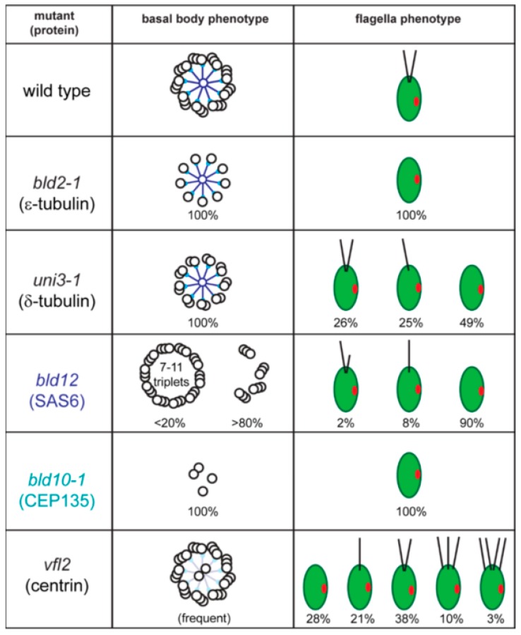
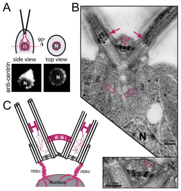
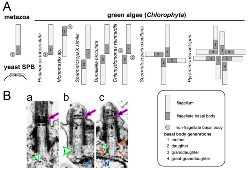
During ciliogenesis, centrioles convert to membrane-docked basal bodies, which initiate the formation of cilia/flagella and template the nine doublet microtubules of the flagellar axoneme. The discovery that many human diseases and developmental disorders result from defects in flagella has fueled a strong interest in the analysis of flagellar assembly. Here, we will review the structure, function, and development of basal bodies in the unicellular green alga Chlamydomonas reinhardtii, a widely used model for the analysis of basal bodies and flagella. Intraflagellar transport (IFT), a flagella-specific protein shuttle critical for ciliogenesis, was first described in C. reinhardtii. A focus of this review will be on the role of the basal bodies in organizing the IFT machinery.

Keywords: axoneme; bld10; bld12; bld2; central pair; centrin; centriole; intraflagellar transport (IFT); microtubules; striated fiber assemblin (SFA).


Conflict of interest statement

The authors declare that they have no conflict of interest.

Figures

Figure 1
Figure 1
Structure of C. reinhardtii flagella and flagellar basal apparatus. (A) Cell overview. The dashed red box indicates the region shown in detail. Schematic drawing of a longitudinal section of a flagellum; the A-, B-, and C-tubules of the basal body are indicated. Red-dashed lines indicate the planes of the TEM cross-sections (ae). (a) Flagellar tip with a mixture of singlet and doublet microtubules indicative for an oblique section. Arrow points to an IFT train. (b) 9 + 2 axoneme surrounded by the plasma membrane and glycocalyx. (c) Transition zone (TZ) with stellate structure, which appears as an H-shaped structure in longitudinal sections (see Figure 3B). The arrow marks a Y-linker. (d) The distal end of the basal body with the transitional fibers (arrows) connecting the triplets to the plasma membrane. (e) Triplet microtubules of the basal body. Arrow: A-C linker. Scale bars = 100 nm. (B) Cell overview, the red dashed line designates the plane of the cross-section. Top: Drawing of the flagellar basal apparatus with the flagella-bearing mother (BB1) and daughter (BB2) basal bodies, the probasal bodies (proBB3) and the microtubular roots consisting of 2 (2MT) or 4 microtubules (4MT). Green striated triangles: SFA fibers. The four fibers are interconnected as indicated by the green shaded area. The 4-stranded root of the daughter basal body connects to the eyespot; it is associated with Mlt1 protein, which participates in eyespot positioning. Bottom: TEM section of the flagellar basal apparatus. Arrows, probasal bodies; arrowheads, microtubular roots.
Figure 2
Figure 2
C. reinhardtii basal body mutants. Wild-type cells possess two flagella and the basal body consist of nine triplet microtubules and a central cartwheel. The spokes of the cartwheel consist of Bld12p/SAS6 (dark blue) and the pinheads of the cartwheel are composed of Bld10p/CEP135 (turquoise). In bld2-1, mutants of ε-tubulin, basal bodies consist of a ring of singlet microtubules and flagella are absent. uni3-1 cells, mutant for δ-tubulin, build a basal body consisting of nine doublet microtubules, and possess 0, 1, or 2 flagella. bld12 cells, mutant for SAS6, possess basal bodies with 8–11 triplets and other defects and mostly lack flagella. A portion of the bld12 cells will build a single flagellum or a pair of unequal length flagella. bld10-1 cells, mutant for Cep135, essentially lack basal bodies and flagella. Centrin RNAi and vfl2 cells lack the stellate structure of the TZ causing the central pair to enter the basal body; they also have defects in basal body duplication and segregation. Values for the distribution of cells with 0–4 flagella are based on Kuchka and Jarvik, 1982.
Figure 3
Figure 3
Distribution of centrin in C. reinhardtii. (A) top: Schematic presentation of cells with the nucleus and the centrin-based nucleus-basal body connectors (NBBCs) (mangenta). Bottom: anti-centrin staining in top (right) and side (left) view. The top view is a focal plane at the level of the nucleus showing the branches of the NBBCs. (B) An electron micrograph (EM) showing a longitudinal-section of the flagella, transition zones (closed arrows), and basal bodies. Open arrows point to the NBBCs. N, nucleus; open arrowhead, distal connecting fiber. The insert (bottom) shows the distal connecting fiber at higher magnification. (C) Cartoon of the centrin-based cytoskeleton (in magenta) of C. reinhardtii. See reference [58] for an outstanding analysis of the distribution of centrin in C. reinhardtii at the ultrastructural level.
Figure 4
Figure 4
Basal body number and maturation in green algae. (A) The mammalian centrosome consists of one mother centriole (1), which might bear a primary cilium, and a daughter basal body (2); a new generation of basal bodies (3) is formed in S phase. Saccharomyces cerevisiae possess a single spindle pole body (SPB) corresponding to one generation of a centrosomal organizer. By comparison green algal centrosomes are more variable with respect to the number of basal bodies, flagella development. (B) TEM images of basal bodies of the green alga S. similis. In mature basal bodies (a), the cartwheel typically consists of three tiers embedded into the axonemal cylinder (green arrowhead). In premitotic cells, the No. 2 basal bodies (b,c) dock to the plasma membrane and develop a protruding cartwheel of ~8 tiers that touches the nuclear envelope (N). Microtubules radiate from the protruding cartwheel suggesting a microtubule organizing center (MTOC) activity. Note the developing TZ. Reprinted with permission from Lechtreck and Grunow (1999) [83].
Figure 5
Figure 5
Basal bodies as organizer for IFT. (A) Overview and total internal reflection fluorescence (TIRF) image of a live cell expressing mNeonGreen-IFT54. The basal body pools (arrows) and the flagellar tips are marked. (B) Series of still images depicting the departure of an anterograde IFT train (arrow) from the basal body pool. T indicates the time in ms. (C) Kymogram (plot of time vs. position) showing the departure and arrival of IFT trains at the flagellar base. Turquoise arrowheads, anterograde trains; pink arrowheads, retrograde trains. Note changes in the signal strength of the basal body pools as trains exit or arrive. (D) Kymogram of IFT traffic in the two flagella. The flagellar base and tip, anterograde trains (turquoise) and retrograde trains (pink) are indicated. Bars (C,D) = 2 μm 2 s. (E) Still images of live cells expressing IFT140-sfGFP (IFT-A; green), NG-IFT20 (IFT-B, red), KAP-GFP (anterograde motor; turquoise) and D1bLIC-GFP (retrograde motor; purple), each of which pool near the two flagella-bearing basal bodies. 10-frame average images are shown for clarity. Bar = 1 μm. (F) Schematic presentation of the distribution of IFT proteins shown in E in the pool in top and side view based on focal series. (G) Model of IFT train assembly. IFT precursor complexes are recruited from the large cell body pool to the basal bodies and assembled into trains by sequential addition of the distinct subcomplexes; complete trains will enter the flagellum. Panels BD are reprinted in modified form Wingfield et al. 2017 [148].

References

    1. Satir P. CILIA: Before and after. Cilia. 2017;6:1. doi: 10.1186/s13630-017-0046-8. - DOI - PMC - PubMed
    1. Brown J.M., Witman G.B. Cilia and Diseases. Bioscience. 2014;64:1126–1137. doi: 10.1093/biosci/biu174. - DOI - PMC - PubMed
    1. Fawcett D.W., Porter K.R. A study of the fine structure of ciliated epithelia. J. Morphol. 1954;94:221–281. doi: 10.1002/jmor.1050940202. - DOI
    1. Musgrave A., de Wildt P., van Etten I., Pijst H., Scholma C., Kooyman R., Homan W., van den Ende H. Evidence for a functional membrane barrier in the transition zone between the flagellum and cell body of Chlamydomonas eugametos gametes. Planta. 1986;167:544–553. doi: 10.1007/BF00391231. - DOI - PubMed
    1. Dentler W.L. Structures linking the tips of ciliary and flagellar microtubules to the membrane. J. Cell Sci. 1980;42:207–220. - PubMed

LinkOut - more resources