. 2018 Jul 17;9(1):2784.
doi: 10.1038/s41467-018-04998-9.

Injectable antibacterial conductive nanocomposite cryogels with rapid shape recovery for noncompressible hemorrhage and wound healing

Affiliations

Injectable antibacterial conductive nanocomposite cryogels with rapid shape recovery for noncompressible hemorrhage and wound healing

Xin Zhao et al. Nat Commun. .

Abstract

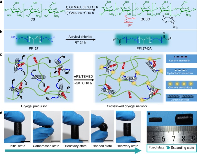
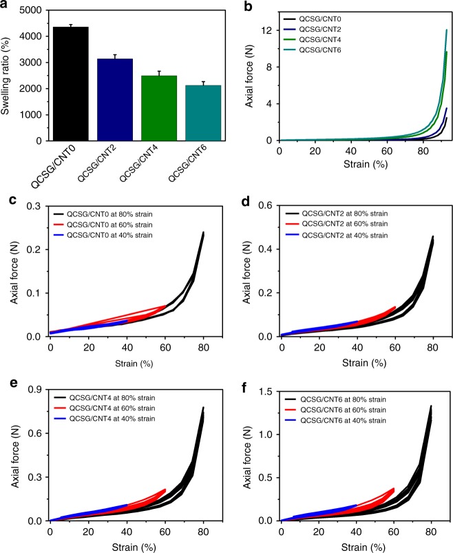
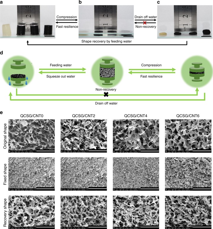
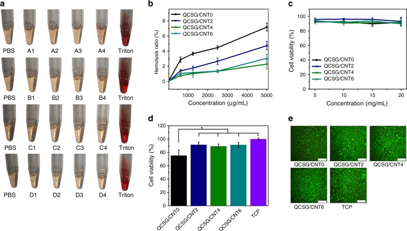
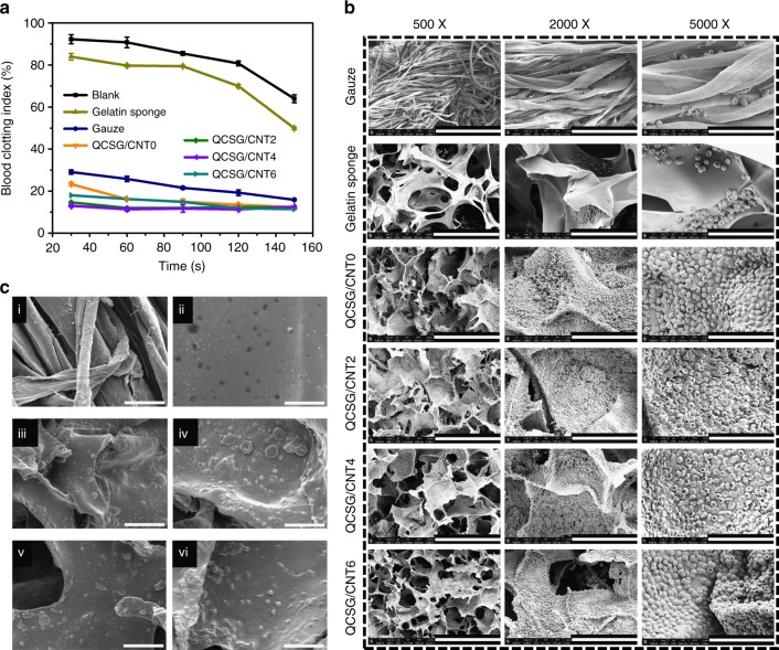
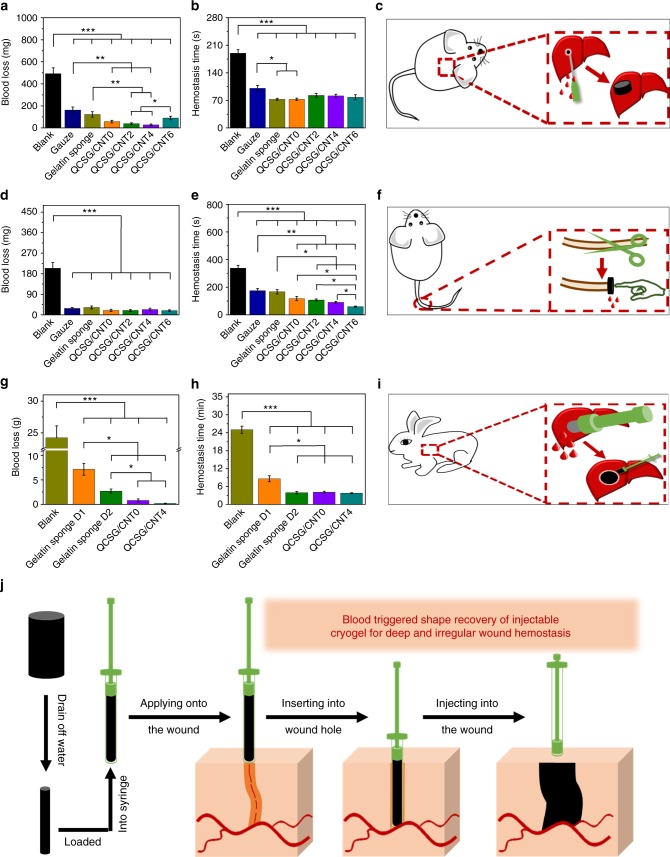
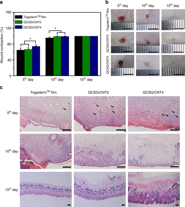
Developing injectable antibacterial and conductive shape memory hemostatic with high blood absorption and fast recovery for irregularly shaped and noncompressible hemorrhage remains a challenge. Here we report injectable antibacterial conductive cryogels based on carbon nanotube (CNT) and glycidyl methacrylate functionalized quaternized chitosan for lethal noncompressible hemorrhage hemostasis and wound healing. These cryogels present robust mechanical strength, rapid blood-triggered shape recovery and absorption speed, and high blood uptake capacity. Moreover, cryogels show better blood-clotting ability, higher blood cell and platelet adhesion and activation than gelatin sponge and gauze. Cryogel with 4 mg/mL CNT (QCSG/CNT4) shows better hemostatic capability than gauze and gelatin hemostatic sponge in mouse-liver injury model and mouse-tail amputation model, and better wound healing performance than Tegaderm™ film. Importantly, QCSG/CNT4 presents excellent hemostatic performance in rabbit liver defect lethal noncompressible hemorrhage model and even better hemostatic ability than Combat Gauze in standardized circular liver bleeding model.


Conflict of interest statement

The Xi’an Jiaotong University has applied for a patent for the discussed hemostatic materials with B.L.G., X.Z., and J.Q. listed as the inventors. The remaining authors declare no competing interests.

Figures

Fig. 1
Fig. 1
Schematic representation of QCSG/CNT cryogel synthesis. a Synthesis of QCSG copolymer. GTMAC and GMA with a fixed 0.5:1 molar ratio of GMA to amino groups and varying the GTMAC: amino groups from 1:1 (coded as QCSG1) to 2:1 (coded as QCSG2) and 3:1 (coded as QCSG3). b Synthesis of PF127-DA copolymer. c Preparation of QCSG/CNT cryogel. d Photographs of the compression and bending resistance capability of QCSG/CNT4 cryogels: initial state, compressed state by squeezing out of the free water, recovery state by absorbing water, bending and squeezing out of part free water, and recovery state after absorbing water. e Shape-fixed state after removing the free water (left) and expanding state after absorbing water (right). Scale bar: 1 cm
Fig. 2
Fig. 2
Swelling and mechanical property of the cryogels. a Swelling ratios of the cryogels. b The uniaxial compression stress–strain curves of the cryogels. QCSG/CNT0 presented the lowest axial force of 2.5 N when bearing a 93% compression strain, while the axial forces significantly increased from 3.5 to 9.6 and 12.0 N with the increase of CNT content from 2 mg/mL to 6 mg/mL in the cryogel networks. Furthermore, all of the four cryogels kept stable and intact after the test. The stress–strain cycling curves of QCSG/CNT0 (c), QCSG/CNT2 (d), QCSG/CNT4 (e), and QCSG/CNT6 (f) with three different compression strains of 40%, 60%, and 80%, respectively. Error bar indicates s.d. (n = 3)
Fig. 3
Fig. 3
Conductivity, photothermal property, release behavior, and antibacterial activity of the cryogels. a Conductivity of the cryogels at wet state and dry state. b ΔT-NIR irradiation time curves of the cryogels using a constant light intensity of 1.4 W/cm2. c ΔT-NIR irradiation time curves of QCSG/CNT4 varying the light intensity from 0.6 to 0.9, 1.1 and 1.4 W/cm2, respectively. d Spontaneous release profiles and NIR-triggered release profiles of ibuprofen from QCSG/CNT0 and QCSG/CNT4. Both QCSG/CNT0 with and without NIR irradiation and QCSG/CNT4 without NIR irradiation  showed similar sustained release profiles as long as 111 h in PBS. However, QCSG/CNT4 presented obvious burst release after applying 10 min NIR irradiation at each time point and completely released the drug within 74 h. When stopping the irradiation, ibuprofen’s release profile returned to its common slow pattern. The killing-time curves of e S. aureus, g E. coli, and i P. aeruginosa for the cryogel groups and PBS group after exposed to NIR irradiation (1.4 W/cm2) for 0 min, 1 min, 3 min, 5 min, 10 min, and 20 min, respectively. Photographs of the survival f S. aureus, h E. coli, and j P. aeruginosa for the cryogel groups and PBS group after exposed to NIR irradiation (1.4 W/cm2) for 0 min, 1 min, 3 min, 5 min, 10 min, and 20 min, respectively. Scale bar: 1 cm. **P < 0.01 using Student's t-test (two-sided). Error bar indicates s.d. (n = 3)
Fig. 4
Fig. 4
Shape memory properties of the cryogels. a–c Fast resilience and macroscopical shape memory property of the cryogels. Scale bar: 1 cm. d Schematic representation of the shape memory mechanism of the cryogel. e Microtopography of the cryogels in original state, shape-fixed state and shape recovery state after fixing. Scale bar: 400 μm. All the four cryogels under free shape showed interconnected macroporous structure with similar pore size between 100–200 µm. Compared to their shapes under free situation, all the shape-fixed cryogels presented collapsed and almost closed pores except QCSG/CNT6 still remained unclosed pores with reduced pore size. However, all the four shape-fixed cryogels still kept unbroken network. After absorbing water, all the cryogels’ morphologies were similar to those in original state
Fig. 5
Fig. 5
Biological properties assays for the cryogels. a Photographs from hemolytic activity assay of the cryogels using PBS as negative control and Triton X-100 as positive control. A: QCSG/CNT0, B: QCSG/CNT2, C: QCSG/CNT4, and D: QCSG/CNT6, and the number after the letter stands for the cryogel dispersion liquid concentration that 1 represents 625 µg/mL, 2 represents 1250 µg/mL, 3 represents 2500 µg/mL, and 4 represents 5000 µg/mL, respectively. b Hemolytic percentage of the cryogels’ dispersion liquids at different concentrations. When the cryogel dispersion concentrations were equal to or less than 1250 µg/mL, all the three CNT-contained cryogels just presented less than 1.8% hemolysis, which was lower than that of QCSG/CNT0 (3.6% hemolysis). When increasing the dispersion concentration to as high as 5000 µg/mL, the three CNT-contained cryogels just presented no more than 4.8% hemolysis. However, the hemolysis ratio of QCSG/CNT0 reached to 7.2%. c Cytocompatibility evaluation of the cryogels’ extracts for L929 cells. When changing the cryogel extracts’ concentrations from 5 to 10, 15, and 20 mg/mL, all the four cryogels presented more than 90% L929 cell viability compared with TCP control group (P > 0.05). d Cytocompatibility evaluation of the cryogels when contacted with the cryogel disks. e LIVE/DEAD staining of L929 cells after contacted with the cryogels for 24 h. Scale bar: 200 µm. *P < 0.05 using Student's t-test (two-sided). Error bar indicates s.d. (n = 4)
Fig. 6
Fig. 6
In vitro hemostatic capacity evaluation of the cryogels. a In vitro dynamic whole-blood-clotting evaluation of the cryogels and controls. The blank group without any hemostatic agents showed the slowest blood-clotting speed and the highest BCI (blood-clotting index) after 150 s. Compared with blank group, gelatin sponge group showed more than a 14% decrease in BCI after 150 s (P < 0.05), while gauze group showed significantly deceased BCI compared to gelatin sponge at each time point (P < 0.001). b SEM images of hemocyte adhesion on the cryogels and controls. Scale bar: 300 µm for ×500; Scale bar: 100 µm for ×2000; Scale bar: 40 µm for ×5000. c SEM images of platelet adhesion on the gauze (ci), gelatin hemostatic sponge (cii), QCSG/CNT0 (ciii), QCSG/CNT2 (civ), QCSG/CNT4 (cv), and QCSG/CNT6 (cvi), respectively. Scale bar: 15 µm. The error bars stand for s.e.m. (n = 3)
Fig. 7
Fig. 7
In vivo hemostatic capacity evaluation of the cryogels. Blood loss (a) and hemostatic time (b) in the mouse liver injury model. The blank group showed the highest blood loss of 492 mg than the other groups (P < 0.001). Gauze and gelatin sponge, as two control groups, presented much decreased blood loss of about 163 mg and 123 mg, respectively, when compared to blank group (P < 0.001). However, all the four cryogels except for QCSG/CNT6 showed significantly decreased blood loss of 57 mg, 38 mg, and 27 mg than those of gauze and the cryogel QCSG/CNT2 and cryogel QCSG/CNT4 also showed significantly decreased blood loss than that of gelatin sponge (P < 0.05). c Scheme representation of the mouse liver injury model during hemostasis. Blood loss (d) and hemostatic time (e) in the mouse-tail amputation model. The blank group (337 s) showed the longest hemostatic time than other groups (P < 0.001). All the four cryogels showed shorter hemostatic times than gauze group (176 s) (P < 0.01), while all the four cryogels except for QCSG/CNT0 showed shorter hemostatic times than gelatin sponge group (167 s) (P < 0.05). f Scheme representation of the mouse-tail amputation model during hemostasis; Blood loss (g) and hemostatic time (h) in the rabbit liver defect lethal noncompressible hemorrhage model. i Scheme representation of the rabbit liver defect lethal noncompressible hemorrhage model during hemostasis. j Scheme representation of the hemostatic application of injectable shape memory cryogel hemostatic in a deep and irregularly shaped wound model. *P < 0.05, **P < 0.01, ***P < 0.001 using Student's t-test (two-sided). The error bars stand for s.e.m. (n = 10 for Fig. 7a, b, d, e; n = 5 for Fig. 7g, h)
Fig. 8
Fig. 8
In vivo wound healing performance of the cryogels. a Wound contraction for TegadermTM film, QCSG/CNT0 and QCSG/CNT4. b Photographs of wounds at 5th, 10th, and 15th day for TegadermTM film, QCSG/CNT0 and QCSG/CNT4. Scale bar: 5 mm. c Histomorphological evaluation of wound regeneration for TegadermTM film, QCSG/CNT0 and QCSG/CNT4 at 5th, 10th, and 15th day. Smooth and complete epithelium layer was presented in two cryogel groups at 10th day, differing from the wound sites in Tegaderm™ group whose epithelium layer was still incomplete and rough. The wounds in QCSG/CNT4 group had a better formation of hair follicles. All wounds were completely healed and characterized with perfect epithelization at 15th day. *P < 0.05 using Student's t-test (two-sided). Scale bar: 5 mm. Error bar indicates s.e.m. (n = 5)

References

    1. Johnson D, et al. The effects of QuikClot Combat Gauze on hemorrhage control in the presence of hemodilution and hypothermia. Ann. Med Surg. 2014;3:21–25. doi: 10.1016/j.amsu.2014.03.001. - DOI - PMC - PubMed
    1. Hee Park D, et al. In vitro degradation and cytotoxicity of alkyl 2‐cyanoacrylate polymers for application to tissue adhesives. J. Appl. Polym. Sci. 2003;89:3272–3278. doi: 10.1002/app.12452. - DOI
    1. Wen J, Weinhart M, Lai B, Kizhakkedathu J, Brooks DE. Reversible hemostatic properties of sulfabetaine/quaternary ammonium modified hyperbranched polyglycerol. Biomaterials. 2016;86:42–55. doi: 10.1016/j.biomaterials.2016.01.067. - DOI - PubMed
    1. Ong SY, Wu J, Moochhala SM, Tan MH, Lu J. Development of a chitosan-based wound dressing with improved hemostatic and antimicrobial properties. Biomaterials. 2008;29:4323–4332. doi: 10.1016/j.biomaterials.2008.07.034. - DOI - PubMed
    1. Revelli L, Tempera SE, Bellantone C, Raffaelli M, Lombardi CP. Minimally Invasive Therapies for Endocrine Neck Diseases. Switzerland: Springer; 2016. pp. 249–259.

Publication types

MeSH terms