. 2018 Jul 17;8(1):10795.
doi: 10.1038/s41598-018-29108-z.

Proteomic Analysis and Functional Characterization of P4-ATPase Phospholipid Flippases from Murine Tissues

Affiliations

Proteomic Analysis and Functional Characterization of P4-ATPase Phospholipid Flippases from Murine Tissues

Jiao Wang et al. Sci Rep. .

Abstract

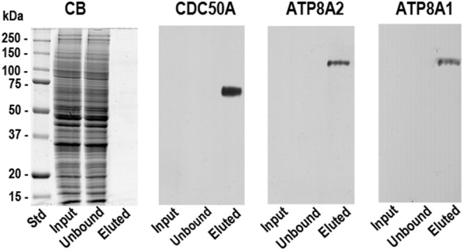
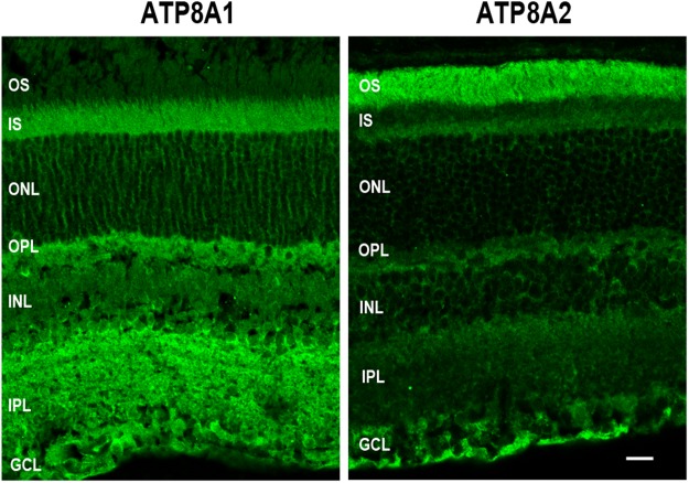
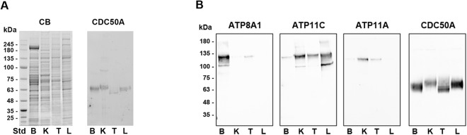
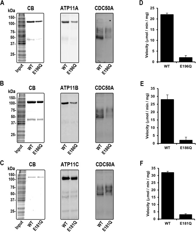
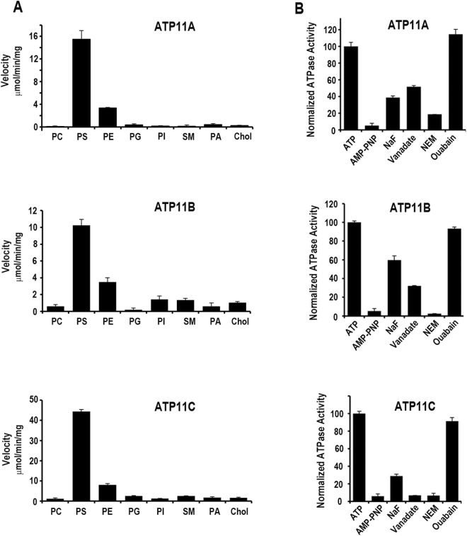
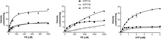
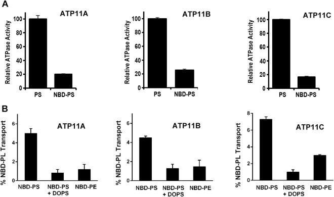
P4-ATPases are a subfamily of P-type ATPases that flip phospholipids across membranes to generate lipid asymmetry, a property vital to many cellular processes. Mutations in several P4-ATPases have been linked to severe neurodegenerative and metabolic disorders. Most P4-ATPases associate with one of three accessory subunit isoforms known as CDC50A (TMEM30A), CDC50B (TMEM30B), and CDC50C (TMEM30C). To identify P4-ATPases that associate with CDC50A, in vivo, and determine their tissue distribution, we isolated P4-ATPases-CDC50A complexes from retina, brain, liver, testes, and kidney on a CDC50A immunoaffinity column and identified and quantified P4-ATPases from their tryptic peptides by mass spectrometry. Of the 12 P4-ATPase that associate with CDC50 subunits, 10 P4-ATPases were detected. Four P4-ATPases (ATP8A1, ATP11A, ATP11B, ATP11C) were present in all five tissues. ATP10D was found in low amounts in liver, brain, testes, and kidney, and ATP8A2 was present in significant amounts in retina, brain, and testes. ATP8B1 was detected only in liver, ATP8B3 and ATP10A only in testes, and ATP8B2 primarily in brain. We also show that ATP11A, ATP11B and ATP11C, like ATP8A1 and ATP8A2, selectively flip phosphatidylserine and phosphatidylethanolamine across membranes. These studies provide new insight into the tissue distribution, relative abundance, subunit interactions and substrate specificity of P4-ATPase-CDC50A complexes.


Conflict of interest statement

The authors declare no competing interests.

Figures

Figure 1
Figure 1
Co-immunoprecipitation of ATP8A2 and ATP8A1 with CDC50A from mouse retina. A retinal extract from mouse retina tissue solubilized in CHAPS detergent was incubated with the immunoaffinity matrix consisting of the Cdc50-7F4 monoclonal antibody covalently coupled to Sepharose 2B. After removal of the unbound fraction, the matrix was washed and eluted with the 7F4 peptide for analysis on SDS gels stained with Coomassie Blue (CB) and western blots labeled with antibodies to CDC50A, ATP8A2 and ATP8A1. The input and unbound lanes contained about 30 µg of protein. Although the P4-ATPases were not detected by Coomassie blue staining due to their low abundance, the ATP8A2, ATP8A1 and CDC50A could be readily detected in the eluted fraction by western blotting.
Figure 2
Figure 2
Immunofluorescence localization of ATP8A1 and ATP8A2 in the mouse retina. Cryosections of adult mouse retina were labeled with ATP8A1 and ATP8A2 antibodies for analysis by confocal scanning microscopy. ATP8A1 and ATP8A2 exhibit some differences in cellular and subcellular localization. ATP8A2 is predominantly present in photoreceptor outer segments and ganglion cell layers, but also present in at a lower level in the inner segments, outer plexiform layer and inner retina. ATP8A1 is absent from outer segments but otherwise displays a more universal distribution throughout the retina. OS:outer segment; IS:inner segment; ONL:outer nuclear layer; OPL:outer plexiform layer; INL:inner nuclear layer; IPL:inner plexiform layer; GCL:ganglion cell layer. Bar = 20 µm
Figure 3
Figure 3
SDS gels and Western blots of P4-ATPase complexes from brain, kidney, testes, and liver. (A) Proteins from membrane fractions of mouse brain (B), kidney (K), testes (T), and liver (L) were resolved on a SDS gel and either stained with Coomassie Blue (CB) or transferred to Immobilon membranes and labeled for CDC50A with the Cdc50-7F4 monoclonal antibody. Approximately, 30 µg of protein was applied to each lane. (B) Western blots of P4-ATPases in mouse tissues isolated by immunoaffinity chromatography. Membranes from brain (B), kidney (K), testes (T), and liver (L) were solubilized in CHAPS detergent and P4-ATPase complexes were isolated by immunoaffinity chromatography on a Cdc50-7F4 immunoaffinity matrix. Western blots were labeled with antibodies to ATP8A1, ATP11C, ATP11A and CDC50A.
Figure 4
Figure 4
Purification and ATPase activity of WT ATP11 and mutants with the E→Q mutation in the activator domain. ATP11A, ATP11B, and ATP11C containing a 1D4 tag were co-expressed with CDC50A in HEK293 cells and purified by immunoaffinity chromatography on a Rho1D4-Sepharose matrix. SDS gels and Western blots of the HEK293 cell extracts (Input) and 1D4 peptide eluted WT or E→Q mutants for ATP11A (A), ATP11B (B) and ATP11C (C). SDS gels were stained with Coomassie Blue (CB) and Western blots were labeled with the Rho 1D4 antibody (ATP11) or Cdc50-7F4 antibody (CDC50A). ATPase activity for WT and E→Q mutants in the presence of brain polar lipid is shown for ATP11A (D), ATP11B (E), and ATP11C (F). Data shown as the mean ± SD for n = 3.
Figure 5
Figure 5
Effect of specific phospholipids, nucleotides and inhibitors on the ATPase activity of ATP11 proteins. (A) ATP11A, ATP11B and ATP11C were co-expressed with CDC50A in HEK293 cells, purified by immunoaffinity chromatography, and reconstituted with 100% DOPC (PC) or 90% DOPC and 10% DOPS (PS), DOPE (PE), DOPG (PG), DOPI (PI), sphingomyelin (SM), DOPA (PA) or cholesterol (Chol) for determination of their ATPase activity. (B) The ATPase activities were determined for 0.5 mM ATP or 0.5 mM non-hydrolyzable ATP analogue AMP-PNP. For inhibition studies, the proteoliposomes were pre-incubated with 100 μM NaF, 100 μM vanadate, 5 mM N-ethylmaleimide (NEM), or 1 mM ouabain prior to the addition of 0.5 mM ATP for ATPase measurements. The ATPase activity was normalized to the activity of proteoliposomes in the presence of ATP, but in the absence of inhibitors. Data shown as the mean ± SD for n = 3.
Figure 6
Figure 6
The effect of PS, PE and ATP concentration on the ATPase activity of purified ATP11A-CDC50A, ATP11B-CDC50A, and ATP11C- CDC50A complexes isolated from transfected HEK293 cells. The purified proteins were reconstituted with DOPC as the base phospholipid and varying concentrations of DOPS (A) or DOPE (B) and the ATPase assays were carried out with 5 mM ATP. (C) The effect of ATP concentration on the ATPase activity for ATP11A, ATP11B and ATP11C complexes reconstituted with 70% DOPC and 30% DOPS. Measurements were performed in triplicate and results were averaged. Error bars represent ± SD. Curves were fitted with a Michaelis-Menten equation using the parameters summarized in Table 2.
Figure 7
Figure 7
ATPase activity and phospholipid flippase activity of ATP11-CDC50A complexes. (A) ATPase activity of ATP11A, ATP11B and ATP11C in the presence of 90% DOPC and either 10% DOPS (PS) or 10% NBD-PS. Activity is normalized to 100% ATPase activity in 10% DOPS. (B) NBD-phospholipid flippase activity. ATP11A, ATP11B and ATP11C associated with CDC50A were purified and reconstituted into DOPC liposomes containing 2.5% NBD-PS, 2.5% NBD-PE or 2.5% NBD-PS plus 30% DOPS. The activity was normalized to samples containing NBD-PS. Addition of 30% unlabeled DOPS effectively competed with NBD-PS to reduce the NBD-PS flipping. Data is the average of 3 experiments ± SD.

References

    1. Andersen JP, et al. P4-ATPases as Phospholipid Flippases-Structure, Function, and Enigmas. Front Physiol. 2016;7:275. doi: 10.3389/fphys.2016.00275. - DOI - PMC - PubMed
    1. Coleman JA, Quazi F, Molday RS. Mammalian P4-ATPases and ABC transporters and their role in phospholipid transport. Biochim Biophys Acta. 2013;1831:555–574. doi: 10.1016/j.bbalip.2012.10.006. - DOI - PMC - PubMed
    1. Lopez-Marques RL, Theorin L, Palmgren MG, Pomorski TG. P4-ATPases: lipid flippases in cell membranes. Pflugers Arch. 2014;466:1227–1240. doi: 10.1007/s00424-013-1363-4. - DOI - PMC - PubMed
    1. Sebastian TT, Baldridge RD, Xu P, Graham TR. Phospholipid flippases: Building asymmetric membranes and transport vesicles. Biochim Biophys Acta. 2012;1821:1068–1077. doi: 10.1016/j.bbalip.2011.12.007. - DOI - PMC - PubMed
    1. Zhu X, et al. Mutations in a p-type ATPase gene cause axonal degeneration. PLoS Genet. 2012;8:e1002853. doi: 10.1371/journal.pgen.1002853. - DOI - PMC - PubMed

Publication types

Substances