. 2018 Jun 26;12(4):042218.
doi: 10.1063/1.5023041. eCollection 2018 Jul.

Barriers-on-chips: Measurement of barrier function of tissues in organs-on-chips

Affiliations

Barriers-on-chips: Measurement of barrier function of tissues in organs-on-chips

Yusuf B Arık et al. Biomicrofluidics. .

Abstract

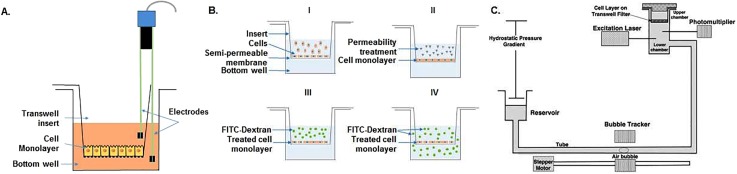
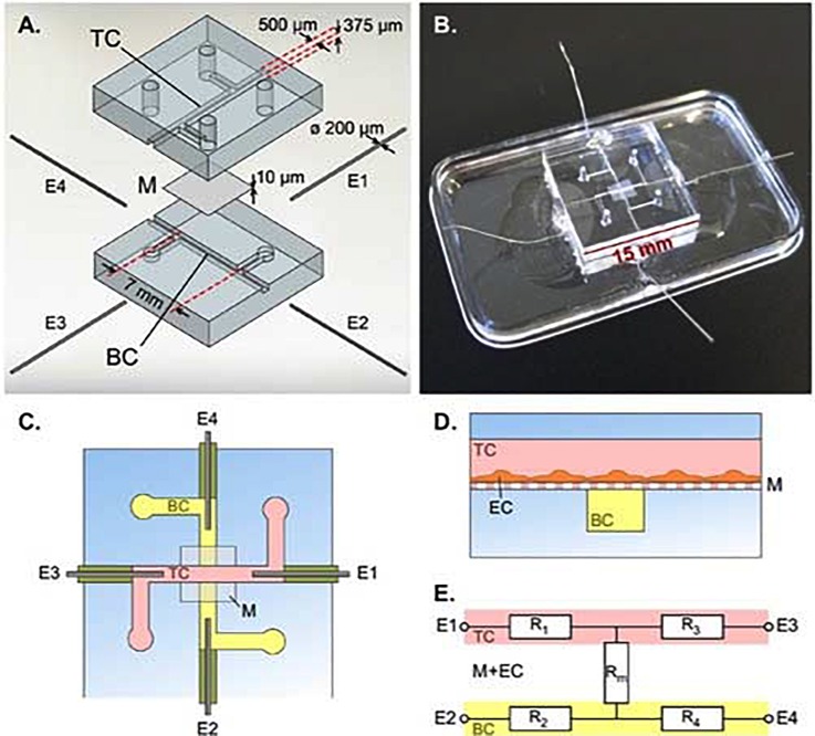
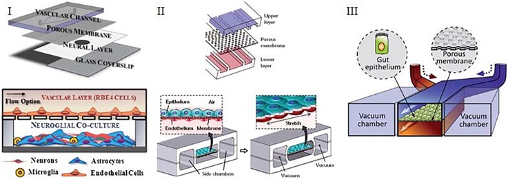
Disruption of tissue barriers formed by cells is an integral part of the pathophysiology of many diseases. Therefore, a thorough understanding of tissue barrier function is essential when studying the causes and mechanisms of disease as well as when developing novel treatments. In vitro methods play an integral role in understanding tissue barrier function, and several techniques have been developed in order to evaluate barrier integrity of cultured cell layers, from microscopy imaging of cell-cell adhesion proteins to measuring ionic currents, to flux of water or transport of molecules across cellular barriers. Unfortunately, many of the current in vitro methods suffer from not fully recapitulating the microenvironment of tissues and organs. Recently, organ-on-chip devices have emerged to overcome this challenge. Organs-on-chips are microfluidic cell culture devices with continuously perfused microchannels inhabited by living cells. Freedom of changing the design of device architecture offers the opportunity of recapitulating the in vivo physiological environment while measuring barrier function. Assessment of barriers in organs-on-chips can be challenging as they may require dedicated setups and have smaller volumes that are more sensitive to environmental conditions. But they do provide the option of continuous, non-invasive sensing of barrier quality, which enables better investigation of important aspects of pathophysiology, biological processes, and development of therapies that target barrier tissues. Here, we discuss several techniques to assess barrier function of tissues in organs-on-chips, highlighting advantages and technical challenges.


Figures

FIG. 1.
FIG. 1.
Conventional barrier assessment methods. (A) TEER measurements in Transwell systems uses two electrode pairs that are submerged in different compartments, measuring resistance of the cellular monolayer seeded onto the membrane. (B) Evaluating barrier by means of transport of fluorescently labeled dextran starts with cells cultured to a monolayer (B-I), then treated with a disease stimulus to change their permeability (B-II). After that FITC-dextran is added to the insert (B-III), and samples can be collected from the bottom compartment to measure the integrity of the cell layer (B-IV). Schematics of experimental setup for measuring hydraulic conductivity (C) starts with Transwell insert sealed in a chamber. For water transport, the reservoir is lowered to create a pressure gradient across the cell layer, then the water flux across the cell monolayer is measured using bubble tracker, and the hydraulic conductivity is determined accordingly [Reproduced with permission from Li et al., Ann. Biomed. Eng. 38, 2499 (2010). Copyright 2010 Springer Nature.]
FIG. 2.
FIG. 2.
Integrated electrodes for measuring TEER in a BBB-Chip. (A) Exploded view of the device with top channel (TC), membrane (M), bottom channel (BC), and platinum wire electrodes (E1, E2, E3, E4). (B) Assembled device. (C) Schematic top view of the device. (D) Cross section schematic of the device showing endothelial cells (EC) cultured on membrane M in the TC. (E) Simplified equivalent circuit of the device, showing electrodes E1-E4, resistors representing the TC (R1 and R3), resistors representing the BC (R2 and R4), and resistor Rm representing the membrane and EC barrier. [Reproduced with permission from van der Helm et al., Biosens. Bioelectron. 85, 924 (2016). Copyright 2016 Elsevier.]
FIG. 3.
FIG. 3.
Typical organ-on-a-chip devices containing two adjacent channels separated by a semi-permeable membrane. (I) Device of Achyuta et al. that consists of 2 parts which are assembled following the cell seeding. [Reproduced with permission from Achyuta et al., Lab Chip 13, 542 (2012). Copyright 2012 The Royal Society of Chemistry.] (II) Device of Huh et al. that consists of two adjacent channels separated by a porous PDMS membrane. [Reproduced with permission from Huh et al., Science 328, 1662 (2010). Copyright 2010 American Association for the Advancement of Science.] (III) Design of Kim et al. that contains two channels, one of which seeded with gut epithelial cells, other containing the interstitial fluid. [Reproduced with permission from Kim et al., Lab Chip 12, 2165 (2012). Copyright 2012 The Royal Society of Chemistry.]
FIG. 4.
FIG. 4.
Microfluidic system reported by Moya et al. for 3D vasculature modelling. (a) PDMS based microfluidic device contains outer microfluidic channels that connect to a series of central micro-tissue chambers through a communication pore on each side. (b) Central chamber is inhabited by endothelial cells and stromal cells embedded in a fibrin matrix [cross-section of panel (a) indicated by a black dotted line]. (c) Hydrostatic pressure is necessary for media flow and is enabled by large media reservoirs. (d) and (e) Microfluidic system enables robust interconnected vessel network formation within 14–21 days (scale bar= 200 μm). [Reproduced with permission from Moya et al., Tissue Eng. Part C 19, 730 (2013). Copyright 2013 Mary Ann Liebert Inc.]

References

    1. Rosenberg G. A., “Neurological diseases in relation to the blood–brain barrier,” J. Cerebral Blood Flow Metabolism 32(7), 1139–1151 (2012). 10.1038/jcbfm.2011.197 - DOI - PMC - PubMed
    1. Cunha-Vaz J. et al. , “Blood-retinal barrier,” Eur. J. Ophthalmol. 21(6_suppl), 3–9 (2011) 10.5301/EJO.2010.6049. - DOI - PubMed
    1. Hurley J., “Current views on the mechanisms of pulmonary oedema,” J. Pathol. 125(2), 59–79 (1978). 10.1002/path.1711250202 - DOI - PubMed
    1. Cucullo L. et al. , “The role of shear stress in Blood-Brain Barrier endothelial physiology,” BMC Neurosci. 12(1), 40 (2011). 10.1186/1471-2202-12-40 - DOI - PMC - PubMed
    1. Huh D. et al. , “Reconstituting organ-level lung functions on a chip,” Science 328(5986), 1662–1668 (2010). 10.1126/science.1188302 - DOI - PMC - PubMed

LinkOut - more resources