Case Reports
. 2019 Feb;27(1):77-83.
doi: 10.1177/1066896918786586. Epub 2018 Jul 18.

ACTG2-Associated Visceral Myopathy With Chronic Intestinal Pseudoobstruction, Intestinal Malrotation, Hypertrophic Pyloric Stenosis, Choledochal Cyst, and a Novel Missense Mutation

Affiliations
Case Reports

ACTG2-Associated Visceral Myopathy With Chronic Intestinal Pseudoobstruction, Intestinal Malrotation, Hypertrophic Pyloric Stenosis, Choledochal Cyst, and a Novel Missense Mutation

Rebecca R J Collins et al. Int J Surg Pathol. 2019 Feb.

Abstract

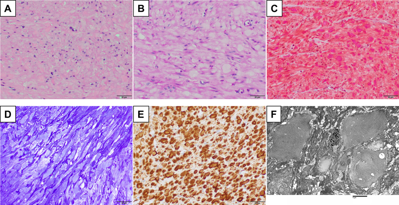
Primary visceral myopathy caused by a pathogenic mutation in the gene encoding the enteric smooth muscle actin gamma 2 ( ACTG2) affects gastrointestinal and genitourinary tracts and often presents as chronic intestinal pseudoobstruction. We present a case of pediatric onset chronic intestinal pseudoobstruction associated with a novel missense ACTG2 mutation c.439G>T/p.G147C. In addition to the known disease manifestations of feeding intolerance and intestinal malrotation, our patient had a late-onset hypertrophic pyloric stenosis and a late-onset choledochal cyst, the former of which has not previously been described in patients with ACTG2-associated visceral myopathy.

Keywords: ACTG2; CIPO; actin gamma 2; choledochal cyst; chronic intestinal pseudoobstruction; hypertrophic pyloric stenosis; visceral myopathy.


Figures

Figure 1.
Figure 1.
A. Abdominal radiograph shows a markedly dilated stomach (yellow arrows). A previously placed gastrojejunostomy tube (red arrow) had retracted into the gastric antrum. B. At laparotomy, a pyloric mass (yellow arrow) and dilated loops of small bowel (red arrows) were noted. C. Gross examination of the resected pyloric mass shows diffuse mural thickening. D. Light microscopic examination shows diffuse smooth muscle hyperplasia consistent with hypertrophic pyloric stenosis; occasional eosinophilic intracytoplasmic irregular inclusions can be seen (hematoxylin and eosin, 400x total magnification). E. Immunohistochemical staining shows smooth muscle actin-positive irregular inclusions in the cytoplasm of smooth muscle cells of the muscularis propria (400x total magnification).
Figure 2.
Figure 2.
Maximum intensity projection from 3D-T2-weighted magnetic resonance cholangiopancreatography shows fusiform dilatation of the common bile duct (red arrow) with dilated intrahepatic biliary ducts (yellow arrow).
Figure 3.
Figure 3.
Photomicrographs of full-thickness duodenal biopsy show haphazardly arranged smooth muscle cells in the muscularis propria with eosinophilic intracytoplasmic irregular inclusions (A. hematoxylin and eosin, 400x total magnification) that do not stain with periodic acid Schiff (B. 400x total magnification) and stain purple with Masson trichrome (C. 400x total magnification) and pale blue with toluidine blue (D. 400x total magnification). The inclusions are smooth muscle actin-positive (E. immunoperoxidase, 400x total magnification) and ultrastructurally composed of irregular aggregates of 9–11 nm filaments (F. 1500x total magnification).
Figure 4.
Figure 4.
A. Sanger sequencing of the entire coding region of the ACTG2 gene identified a novel heterozygous missense mutation c.439G>T that results in replacement of the glycine at position 147 with cysteine. B. A cartoon of the exons (numbered 1–9) and coding region (in gray) of the ACTG2 gene shows the location of the reported mutations associated with visceral myopathy.
Figure 5.
Figure 5.
A. A cartoon representation showing the structural context for the mutated site. Actin (pale blue) and a bound WH2 domain from WASP (tan) from Protein Data Bank entry 2A3Z (https://www.rcsb.org/structure/2A3Z) are shown, with ATP as sticks. The position of the mutated site is indicated with a red sphere. B. A comparison across species shows that the ACTG2 protein sequence including the G147 residue (arrow) is evolutionarily conserved (Alamut Visual, Interactive Biosoftware, Rouen, France).

References

    1. Knowles CH, De Giorgio R, Kapur RP, Bruder E, Farrugia G, Geboes K, et al. The London Classification of gastrointestinal neuromuscular pathology: report on behalf of the Gastro 2009 International Working Group. Gut. 2010;59:882–887. - PubMed
    1. Knowles CH, De Giorgio R, Kapur RP, Bruder E, Farrugia G, Geboes K, et al. Gastrointestinal neuromuscular pathology: guidelines for histological techniques and reporting on behalf of the Gastro 2009 International Working Group. Acta Neuropathol. 2009;118:271–301. - PubMed
    1. Di Nardo G, Di Lorenzo C, Lauro A, Stanghellini V, Thapar N, Karunaratne TB, et al. Chronic intestinal pseudo-obstruction in children and adults: diagnosis and therapeutic options. Neurogastroenterol Motil. 2017;29. - PubMed
    1. Thapar N, Saliakellis E, Benninga MA, Borrelli O, Curry J, Faure C, et al. Paediatric Intestinal Pseudo-Obstruction: Evidence and Consensus-Based Recommendations from an ESPGHAN-Led Expert Group. J Pediatr Gastroenterol Nutr. 2018. - PubMed
    1. Wangler MF, Beaudet AL. ACTG2-Related Disorders. In: Pagon RA, Adam MP, Ardinger HH, et al., eds. GeneReviews(R) Seattle (WA); 2015.

Publication types

LinkOut - more resources