Review
. 2018 Jul-Aug;44(4):321-333.
doi: 10.1590/S1806-37562017000000185. Epub 2018 Jul 6.

Patient-ventilator asynchrony

[Article in English, Portuguese]
Affiliations
Review

Patient-ventilator asynchrony

[Article in English, Portuguese]
Marcelo Alcantara Holanda et al. J Bras Pneumol. 2018 Jul-Aug.

Erratum in

  • ERRATUM.
    [No authors listed] [No authors listed] J Bras Pneumol. 2018 Jul-Aug;44(4):339. doi: 10.1590/S1806-37562017000000185errata. J Bras Pneumol. 2018. PMID: 30183973 Free PMC article.

Abstract

Patient-v entilator asynchrony (PVA) is a mismatch between the patient, regarding time, flow, volume, or pressure demands of the patient respiratory system, and the ventilator, which supplies such demands, during mechanical ventilation (MV). It is a common phenomenon, with incidence rates ranging from 10% to 85%. PVA might be due to factors related to the patient, to the ventilator, or both. The most common PVA types are those related to triggering, such as ineffective effort, auto-triggering, and double triggering; those related to premature or delayed cycling; and those related to insufficient or excessive flow. Each of these types can be detected by visual inspection of volume, flow, and pressure waveforms on the mechanical ventilator display. Specific ventilatory strategies can be used in combination with clinical management, such as controlling patient pain, anxiety, fever, etc. Deep sedation should be avoided whenever possible. PVA has been associated with unwanted outcomes, such as discomfort, dyspnea, worsening of pulmonary gas exchange, increased work of breathing, diaphragmatic injury, sleep impairment, and increased use of sedation or neuromuscular blockade, as well as increases in the duration of MV, weaning time, and mortality. Proportional assist ventilation and neurally adjusted ventilatory assist are modalities of partial ventilatory support that reduce PVA and have shown promise. This article reviews the literature on the types and causes of PVA, as well as the methods used in its evaluation, its potential implications in the recovery process of critically ill patients, and strategies for its resolution.

A assincronia pacie nte-ventilador (APV) é um desacoplamento entre o paciente, em relação a demandas de tempo, fluxo, volume e/ou pressão de seu sistema respiratório, e o ventilador, que as oferta durante a ventilação mecânica (VM). É um fenômeno comum, com taxas de incidência entre 10% e 85%. A APV pode ser devida a fatores relacionados ao paciente, ao ventilador ou a ambos. Os tipos de APV mais comuns são as de disparo, como esforço ineficaz; autodisparo e duplo disparo; as de ciclagem (tanto prematura quanto tardia); e as de fluxo (insuficiente ou excessivo). Cada um desses tipos pode ser detectado pela inspeção visual das curvas de volume-tempo, fluxo-tempo e pressãotempo na tela do ventilador mecânico. Estratégias ventilatórias específicas podem ser adotadas, em combinação com a abordagem clínica do paciente, como controle de dor, ansiedade, febre, etc. Níveis profundos de sedação devem ser evitados sempre que possível. A APV se associa a desfechos indesejados, tais como desconforto, dispneia, piora da troca gasosa, aumento do trabalho da respiração, lesão muscular diafragmática, prejuízo do sono, aumento da necessidade de sedação e/ou de bloqueio neuromuscular, assim como aumento do tempo de VM, de desmame e de mortalidade. A ventilação proporcional assistida e a ventilação assistida com ajuste neural são modalidades de suporte ventilatório parcial que reduzem a APV e têm se mostrado promissoras. Este artigo revisa a literatura acerca da APV abordando seus tipos, causas, métodos de avaliação, suas potenciais implicações no processo de recuperação de pacientes críticos e estratégias para sua resolução.


Figures

Figure 1
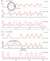
Figure 1. Flow and pressure waveforms, respectively, illustrating two simulated types of ineffective triggering. The first two waveforms represent a patient without problems in respiratory mechanics, with a weak spontaneous effort (Pmus) because of respiratory muscle weakness or decreased neural drive. The bottom two waveforms represent a patient with airflow obstruction and difficulty in triggering some breaths because of the presence of auto-positive endexpiratory pressure, even with a muscle effort that is “physiological” but unable to trigger ventilator breaths. In both cases, pressure-controlled ventilation (pressure sensitivity of −2 cmH2O) was used. The dots on the waveforms indicate ineffective efforts. Paw: airway pressure; and Pmus: muscle pressure. Source: Xlung®.
Figure 2
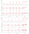
Figure 2. Volume, flow and pressure waveforms, respectively, illustrating two simulated types of auto-triggering. The first three waveforms represent a patient on pressure-support ventilation with flow sensitivity. The system with a leak causes the onset of flow-triggered breaths, without patient effort (Pmus = 0). The bottom three waveforms represent a patient on pressure-controlled ventilation,* without respiratory muscle effort, but showing regular flow and pressure oscillations, with a respiratory rate of approximately 80 breaths/min, corresponding to his/her heart rate. Pressure sensitivity was changed to flow sensitivity. The increase in the total respiratory rate was due to triggers induced by transmission of flow oscillations because of cardiac activity. Vol.: volume; and Paw: airway pressure. Source: Xlung®.
Figure 3
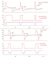
Figure 3. Volume, flow, and pressure waveforms, respectively, illustrating two simulations of asynchrony. The first three waveforms represent a case in which, because of patient neural inspiratory time, which is longer than the ventilator inspiratory time, the first breath is always triggered by the patient, during volume-controlled ventilation. The dots indicate stacked tidal volume caused by double triggering. The bottom three waveforms represent a case of reverse triggering due to respiratory muscle effort triggered by reflex mechanisms resulting from a ventilator-delivered breath, during pressure-controlled ventilation. Note, in both cases, stacked tidal volume and increased airway pressure during asynchrony. The dots indicate reverse triggering. Paw: airway pressure; and Pmus: muscle pressure. Source: Xlung®.
Figure 4
Figure 4. Flow and pressure waveforms, respectively, illustrating two types of cycling asynchrony simulated during pressure support ventilation. The first two waveforms represent a patient with COPD. Asynchrony is corrected by increasing the threshold percentage of peak inspiratory flow for termination of inspiration. The bottom two waveforms represent a patient with restrictive lung disease experiencing premature cycling. Asynchrony is attenuated by decreasing the cycling threshold percentage of peak flow. The dots indicate cycling during pressure support ventilation. Paw: airway pressure; and Pmus: muscle pressure. Source: Xlung®.
Figure 5
Figure 5. Volume, flow, and pressure waveforms, respectively, illustrating simulation of correction of flow asynchrony and volume asynchrony (air hunger), evident in the second breath, during VCV. The application of PCV from the third breath onward enabled delivery of flow and tidal volume. The patient responded with decreased muscle contraction (Pmus) from the fourth breath onward. Note a slight airway pressure overshoot at the end of breath during PCV (arrow), attenuated by better adaptation of the patient. The dots indicate free-flow delivery during PCV. VCV: volume-controlled ventilation; PCV: pressure-controlled ventilation; Paw: airway pressure;; and Pmus: muscle pressure. Source: Xlung®.

References

    1. Wunsch H, Linde-Zwirble WT, Angus DC, Hartman ME, Milbrandt EB, Kahn JM. The epidemiology of mechanical ventilation use in the United States. Crit Care Med. 2010;38(10):1947–1953. doi: 10.1097/CCM.0b013e3181ef4460. - DOI - PubMed
    1. Barbas CS, Isola AM, Farias AM, Cavalcanti AB, Gama AM, Duarte AC. Brazilian recommendations of mechanical ventilation 2013 Part I. Rev Bras Ter Intensiva. 2014;26(2):89–121. doi: 10.5935/0103-507X.20140017. - DOI - PMC - PubMed
    1. Vasconcelos Rdos S, Melo LH, Sales RP, Marinho LS, Deulefeu FC, Reis RC. Effect of an automatic triggering and cycling system on comfort and patient-ventilator synchrony during pressure support ventilation. Respiration. 2013;86(6):497–503. doi: 10.1159/000353256. - DOI - PubMed
    1. Chao DC, Scheinhorn DJ, Stearn-Hassenpflug M. Patient-ventilator trigger asynchrony in prolonged mechanical ventilation. Chest. 1997;112(6):1592–1599. doi: 10.1378/chest.112.6.1592. - DOI - PubMed
    1. Thille AW, Rodriguez P, Cabello B, Lellouche F, Brochard L. Patientventilator asynchrony during assisted mechanical ventilation. Intensive Care Med. 2006;32(10):1515–1522. doi: 10.1007/s00134-006-0301-8. - DOI - PubMed

MeSH terms