

Effects of diode laser application on inflammation and mpo in periodontal tissues in a rat model
- PMID: 30020350
- PMCID: PMC6089569
- DOI: 10.1590/1678-7757-2017-0266
Effects of diode laser application on inflammation and mpo in periodontal tissues in a rat model
Abstract
Objective: In this study, we aimed to histologically and immunologically evaluate the effect of diode laser treatment when applied adjunctive to scaling and root planing (SRP) in an experimental periodontitis model.
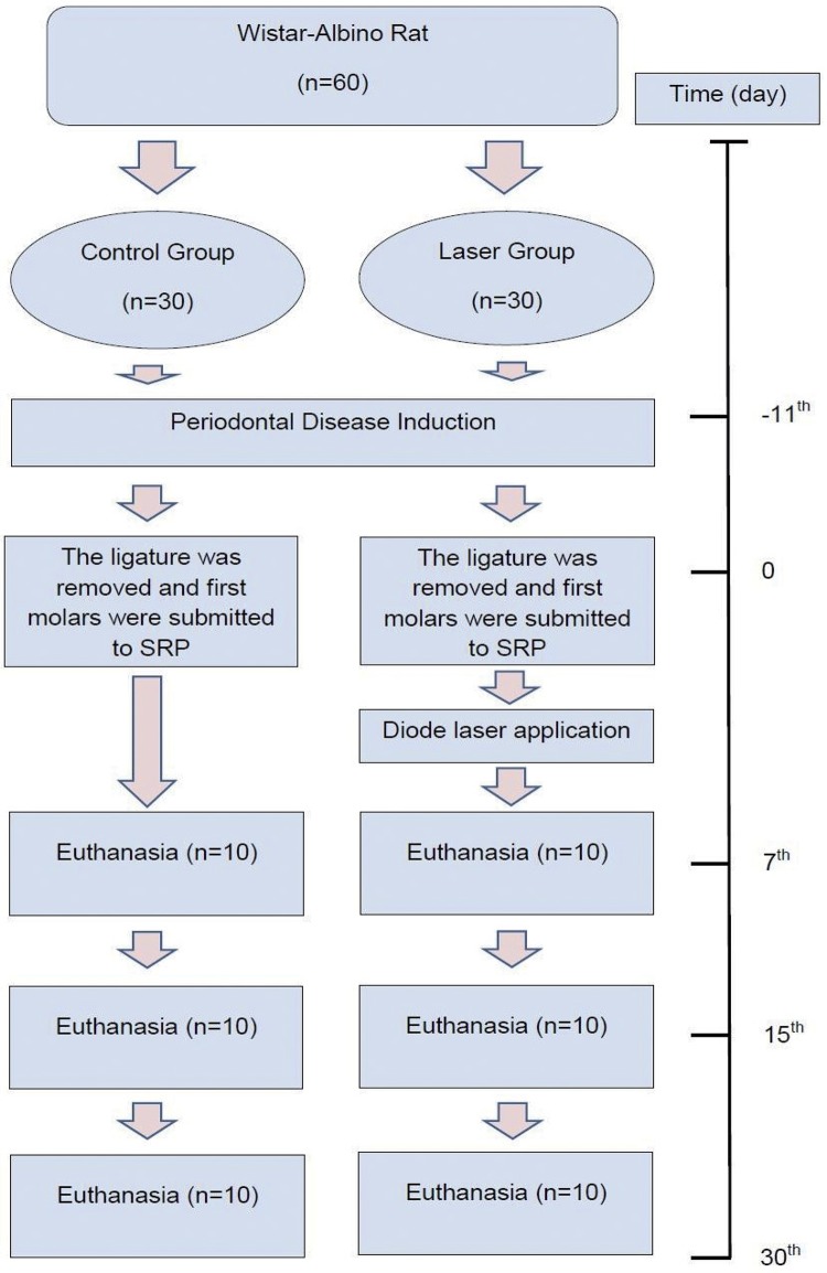
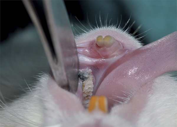
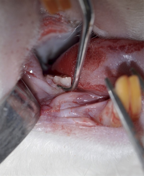
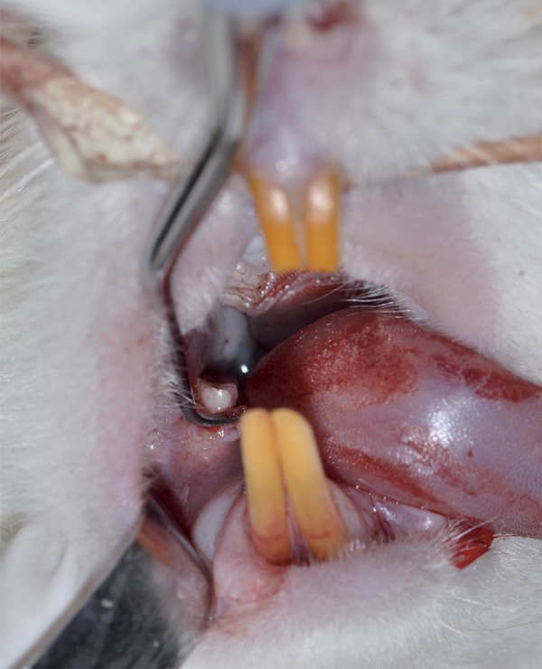
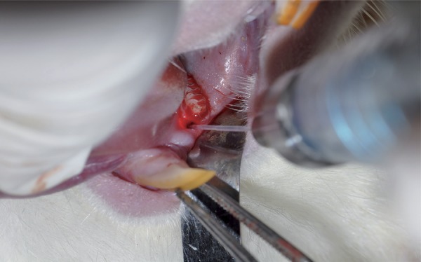
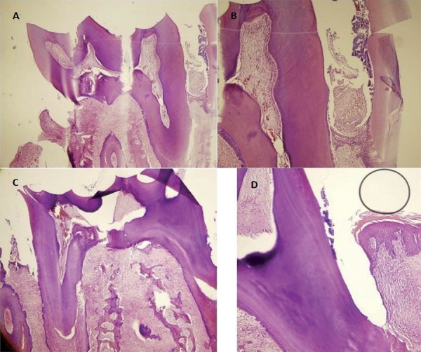
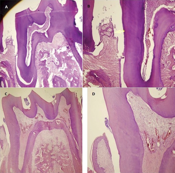
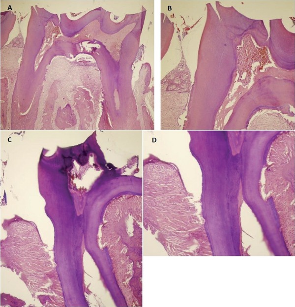
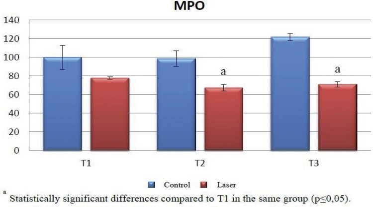
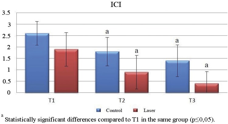
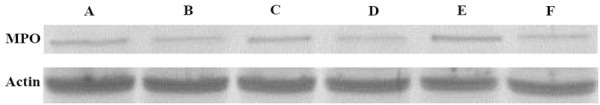
Materials and methods: We used Wistar-Albino rats (n=60) with average weight of 230 g. Experimental periodontitis was induced by ligature at the right and left first mandibular molar teeth in all rats. After 11 days, the ligature was removed and rats were divided into two groups. The control group (n=30) received only SRP treatment, while the laser group (n=30) received a diode laser (GaAlAs, 810 nm, 1 W, 10 J, 20 s) treatment adjunctive to SRP. Ten rats in each group were sacrificed after 7, 15, and 30 days. Histopathological examination was performed in the left mandible of rats. Myeloperoxidase (MPO) was evaluated by western blot in the gingival specimens from the right mandible.
Results: MPO levels in the laser group were statistically significantly lower compared with the control group (p≤0.05). There was no statistically significance at any time between MPO levels in the control group (p>0.05). MPO levels in the laser group at the 7th day were statistically significantly higher compared to the 15th (p≤0.05) and the 30th day (p≤0.05). Inflammatory cell infiltration decreased over time in both groups and was statistically significantly lower in the laser group than in the control group at all times (p≤0.01).
Conclusions: Within the limits of this study, we suggest that diode laser application is an adjunctive treatment because it reduced inflammation and MPO when applied in addition to SRP. On the other hand, more studies are needed for the assessment of the effects of diode laser application to periodontal tissues.
Conflict of interest statement
Disclosure statement
The authors declare no conflict of interest.
Figures
References
-
- Lindhe J, Okamoto H, Yoneyama T, Haffajee A, Socransky SS. Longitudinal changes in periodontal disease in untreated subjects. J Clin Periodontol. 1989;16(10):662–670. - PubMed
-
- Canakci CF, Tatar A, Canakci V, Cicek Y, Oztas S, Orbak R. New evidence of premature oxidative DNA damage: mitochondrial DNA deletion in gingival tissue of patients with periodontitis. J Periodontol. 2006;77(11):1894–1900. - PubMed
-
- Chapple IL, Matthews JB. The role of reactive oxygen and antioxidant species in periodontal tissue destruction. Periodontol 2000. 2007;43:160–232. - PubMed
-
- Leppilahti J, Hernandez-Rios P, Gamonal J, Tervahartiala T, Brignardello-Petersen R, Mantyla P, et al. Matrix metalloproteinases and myeloperoxidase in gingival crevicular fluid provide site-specific diagnostic value for chronic periodontitis. J Clin Periodontol. 2014;41(4):348–356. - PubMed
Publication types
MeSH terms
Substances
LinkOut - more resources
Full Text Sources
Other Literature Sources
Research Materials
Miscellaneous
