

Clinical and Pathological Aspects of Silent Pituitary Adenomas
- PMID: 30020466
- PMCID: PMC6517166
- DOI: 10.1210/jc.2018-00688
Clinical and Pathological Aspects of Silent Pituitary Adenomas
Abstract
Context: Silent pituitary adenomas are anterior pituitary tumors with hormone synthesis but without signs or symptoms of hormone hypersecretion. They have been increasingly recognized and represent challenging diagnostic issues.
Evidence acquisition: A comprehensive literature search was performed using MEDLINE and EMBASE databases from January 2000 to March 2018 with the following key words: (i) pituitary adenoma/tumor and nonfunctioning; or (ii) pituitary adenoma/tumor and silent. All titles and abstracts of the retrieved articles were reviewed, and recent advances in the field of silent pituitary adenomas were summarized.
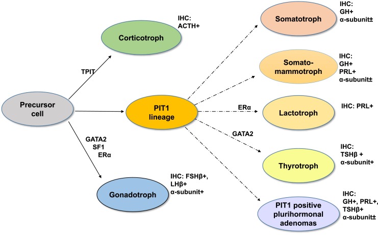
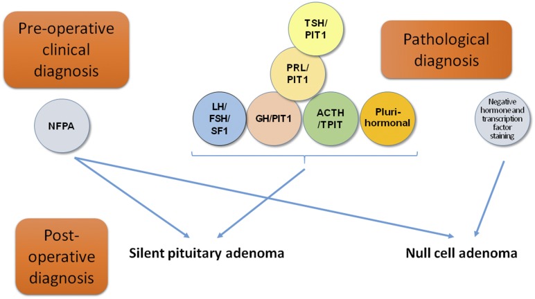
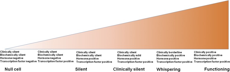
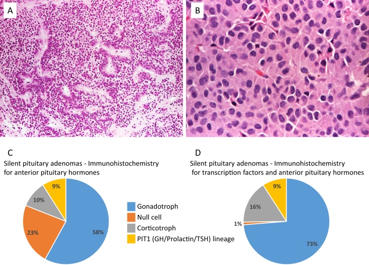
Evidence synthesis: The clinical and biochemical picture of pituitary adenomas reflects a continuum between functional and silent adenomas. Although some adenomas are truly silent, others will show some evidence of biochemical hypersecretion or could have subtle clinical signs and, therefore, can be referred to as clinically silent or "whispering" adenomas. Silent tumors seem to be more aggressive than their secreting counterparts, with a greater recurrence rate. Transcription factors for pituitary cell lineages have been introduced into the 2017 World Health Organization guidelines: steroidogenic factor 1 staining for gonadotroph lineage; PIT1 (pituitary-specific positive transcription factor 1) for growth hormone, prolactin, and TSH lineage, and TPIT for the corticotroph lineage. Prospective studies applying these criteria will establish the value of the new classification.
Conclusions: A concise review of the clinical and pathological aspects of silent pituitary adenomas was conducted in view of the new World Health Organization classification of pituitary adenomas. New classifications, novel prognostics markers, and emerging imaging and therapeutic approaches need to be evaluated to better serve this unique group of patients.
Copyright © 2019 Endocrine Society.
Figures
References
-
- Asa SL, Casar-Borota O, Chanson P, Delgrange E, Earls P, Ezzat S, Grossman A, Ikeda H, Inoshita N, Karavitaki N, Korbonits M, Laws ER Jr, Lopes MB, Maartens N, McCutcheon IE, Mete O, Nishioka H, Raverot G, Roncaroli F, Saeger W, Syro LV, Vasiljevic A, Villa C, Wierinckx A, Trouillas J; Attendees of 14th Meeting of the International Pituitary Pathology Club, Annecy, France, November 2016 . From pituitary adenoma to pituitary neuroendocrine tumor (PitNET): an International Pituitary Pathology Club proposal. Endocr Relat Cancer. 2017;24(4):C5–C8. - PubMed
-
- Daly AF, Rixhon M, Adam C, Dempegioti A, Tichomirowa MA, Beckers A. High prevalence of pituitary adenomas: a cross-sectional study in the province of Liege, Belgium. J Clin Endocrinol Metab. 2006;91(12):4769–4775. - PubMed
-
- Lloyd R, Osamura R, Klöppel G, Rosai J, eds. World Health Organization Classification of Tumours of Endocrine Organs, 4th ed Volume 10 Lyon, France: IARC Publication; 2017.
-
- Mete O, Lopes MB. Overview of the 2017 WHO classification of pituitary tumors. Endocr Pathol. 2017;28(3):228–243. - PubMed
-
- Fernandez A, Karavitaki N, Wass JA. Prevalence of pituitary adenomas: a community-based, cross-sectional study in Banbury (Oxfordshire, UK). Clin Endocrinol (Oxf). 2010;72(3):377–382. - PubMed
Publication types
MeSH terms
Substances
Grants and funding
LinkOut - more resources
Full Text Sources
Other Literature Sources
Medical
