. 2019 Jan;29(1):114-125.
doi: 10.1111/bpa.12644. Epub 2018 Oct 5.

A clinical and histopathological study of malformations observed in fetuses infected by the Zika virus

Affiliations

A clinical and histopathological study of malformations observed in fetuses infected by the Zika virus

Aurélie Beaufrère et al. Brain Pathol. 2019 Jan.

Abstract

Background: The recent outbreak of Zika virus (ZIKV) infection and the associated increased prevalence of microcephaly in Brazil underline the impact of viral infections on embryo fetal development. The aim of the present study is to provide a detailed clinical and histopathological study of the fetal disruption caused by the ZIKV, with a special focus on the associated neuropathological findings.

Methods: A detailed feto-placental examination, as well as neuropathological and neurobiological studies were performed on three fetuses collected after pregnancy termination between 22 and 25 weeks of gestation (WG), because brain malformations associated with a maternal and fetal ZIKV infection was diagnosed.

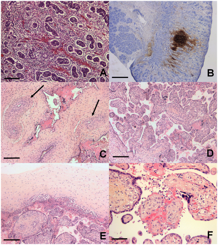
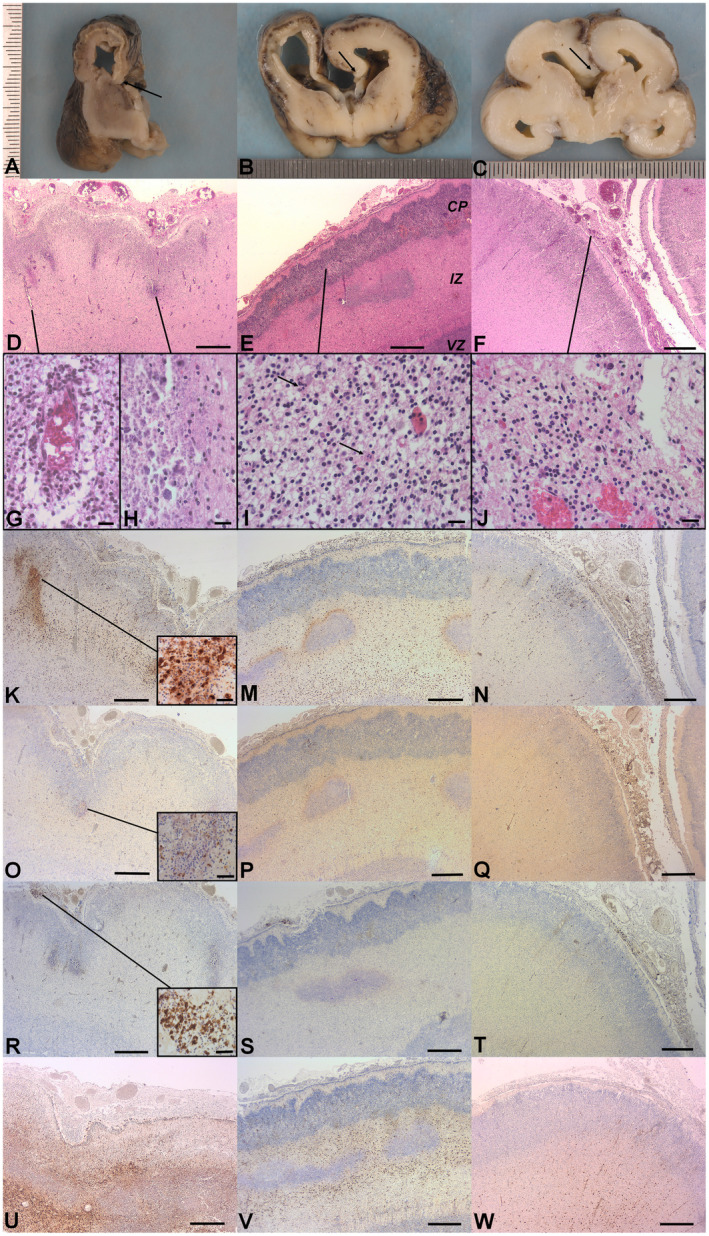
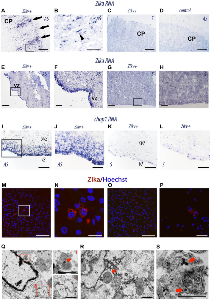
Results: In all three cases, the maternal infection occurred during the first trimester of pregnancy. A small head was observed on the ultrasound examination of the second trimester of pregnancy and led to the diagnosis of ZIKV fetopathy and pregnancy termination. The fetal histopathological examination was unremarkable on the viscera but showed on the testis an interstitial lymphocytic infiltrate. The placenta contained a Hofbauer cells hyperplasia with signs of inflammation. Neuropathological findings included a meningoencephalitis and an ex vacuo hydrocephalus. Immunohistochemical studies showed the presence of T lymphocytic and histiocytic meningitis associated with an abundant cerebral astroglial and macrophagic reaction. In situ hybridization demonstrated, abundant ZIKV particles within the cerebral parenchyma mainly in the ventricular/subventricular zone and in the cortical plate. In addition massive cells death and endoplasmic reticulum damage were present.

Conclusion: The present study reports on the clinical and histopathological findings observed in three fetuses infected by the ZIKV. It emphasizes the severity of brain damages and the minimal visceral and placental changes observed upon ZIKV infection. This confirms the selective neurotropism of ZIKV. Finally, it allows us to describe the cascade of multifactorial developmental defects leading to microcephaly.

Keywords: ER stress; ZIKV infection.; fetus; meningoencephalitis; micrencephaly; microcephaly.


Conflict of interest statement

None.

Figures

Figure 1
Figure 1
Testes and placental analysis. A. Histological section: interstitial lymphocytic infiltrate in testis. B. Anti CD20 immunohistochemistry: interstitial nodular lymphocytic infiltrate in testis. C. Fibromuscular hypertrophy of truncal vessels (arrows). D. Hofbauer cells hyperplasia. E. Acute chorioamnionitis. F. Chronic villitis. H&E, scale bar: 400 μm (B), 80 μm (A, D, E), 40 μm (C, F).
Figure 2
Figure 2
Neuropathological findings. Gross examination. A. Case 1: Coronal section of the right cerebral hemisphere showing: the lateral ventricle dilatation, the thin cerebral mantle, the cortical plate is separated from the underlying intermediate zone by a rim of mineralized tissue, thin corpus callosum (arrow), reduced deep brain nuclei. B. Case 2: Coronal section of cerebral hemispheres showing: triventricular dilatation, short corpus callosum (arrow), diffuse cortical hemorrhagic lesions. C. Case 3: Coronal section of cerebral hemispheres showing: triventricular dilatation and absent corpus callosum (arrow), focal cortical hemorrhagic lesion. Histological findings. D. Case 1: The cerebral mantle (H&E): Note diffuse inflammatory infiltrate in the arachnoid, associated to ependymal lesions and necrotic zones at the interface between the cortical plate (CP) and the intermediate zone (IZ). E. Case 2: The cerebral mantle (H&E): Note diffuse inflammatory infiltrate in the arachnoid, associated to ependymal lesions and necrotic zones at the interface between the CP and the IZ. CP: cortical plate, IZ: intermediate zone, VZ: ventricular zone. F. Case 3: The cerebral mantle (H&E): Note diffuse inflammatory infiltrate in the arachnoid, associated to ependymal lesions. G. Vasculitis in case 1 (high magnification, H&E). H. Calcified necrotic zone in case 1 (high magnification, H&E). I. Cerebral cortex case 2 (high magnification, H&E): Neuronal rarefaction, apoptotic bodies (arrow). J. Case 3: Inflammatory infiltrate in the arachnoid composed of numerous macrophages and lymphocytes (H&E). Immunohistochemistry. K. M. N. Anti CD68: macrophagic reaction in the wall of cerebral hemisphere, mainly around the necrotic zones in case 1 (magnified field) and 2. O. P. Q. Anti CD3: T lymphocytes are less numerous than macrophages but are also found in all cerebral areas and mainly around necrotic zones (magnified field). R. S. T. Anti CD20: few lymphocytes marked in the arachnoid (magnified field). U. V. W. Anti GFAP: hyperplastic and hypertrophic astrocytes in cerebral parenchyma. Scale bar: 400 μm (D, E, F, K, M, N, O, P, Q, R, S, T, U, V, W), 40 μm (G, H, I, J, magnified fields K, O and R).
Figure 3
Figure 3
Histological and cellular analyses of ZIKV‐infected brains. A–L, in situ hybridization on coronal sections from human fetus ZIKV‐infected (Zikv+, case 1 A–C, E–H, and I–L) ZIKV‐uninfected (D,control) with antisense (AS) and sense (S) RNA probes of Zikv (A–H) and chop (I–L). B, F, and H are the magnifications of insets in A, E, and G, respectively. CP, cortical plate; VZ, ventricular zone. The black arrowhead indicates a single infected bipolar neuron migrating to the CP. M–P Immunolabeling of coronal sections from (case 1) ZIKV‐infected fetal brain with an antibody specific for NS1 (non‐structural protein 1 expressed by flaviviruses). M, O, P represent different dorsal regions of the cerebral cortex of case1 ZIKV‐infected fetal brain. N is a magnification of the insets in M. NS1 is in Red and nuclei are labeled by Hoechst (blue). Q–S, transmission electronic micrographs of frontal cortical sample from the brain of the (case 1) ZIKV‐infected fetus showing a cell with an autophagosome with an electron‐dense 50‐nm ZIKV particle (red square 1) and enlarged ER (red square 2) (Q), another one with enlarged ER and autophagosomes with ZIKV particles (R, S). Red arrows point ZIKV particles. Scale bar: 150 μm (A, C, D), 40 μm (B), 250 μm (E, G, H), 50 μm (F, J, L, M, O), 100 μm (I, K), 10μm (N), 20μm (P), 1 μm (Q, left), 500 nm (Q, right; R; S).

References

    1. Aagaard KM, Lahon A, Suter MA, Arya RP, Seferovic MD, Vogt MB et al (2017) Primary human placental trophoblasts are permissive for zika virus (ZIKV) replication. Sci Rep 7:41389. - PMC - PubMed
    1. Benirschke K, Mendoza GR, Bazeley PL (1974) Placental and fetal manifestations of cytomegalovirus infection. Virchows Arch B Cell Pathol 16(2):121. - PubMed
    1. Besnard M, Eyrolle‐Guignot D, Guillemette‐Artur P, Lastère S, Bost‐Bezeaud F, Marcelis L, et al (2016) Congenital cerebral malformations and dysfunction in fetuses and newborns following the 2013 to 2014 Zika virus epidemic in French Polynesia. Euro Surveill Bull Eur Sur Mal Transom Eur Common Dis Bull 21(13). - PubMed
    1. Bhatnagar J, Rabeneck DB, Martines RB, Reagan‐Steiner S, Ermias Y, Estetter LBC, et al (2017) Zika virus RNA replication and persistence in brain and placental tissue. Emerg Infect Dis 23(3):405. - PMC - PubMed
    1. Blázquez A‐B, Saiz J‐C (2016) Neurological manifestations of Zika virus infection. World J Virol 5(4):135–143. - PMC - PubMed

Publication types