. 2018 Nov;77(11):1653-1664.
doi: 10.1136/annrheumdis-2018-213197. Epub 2018 Jul 18.

Photosensitivity and type I IFN responses in cutaneous lupus are driven by epidermal-derived interferon kappa

Affiliations

Photosensitivity and type I IFN responses in cutaneous lupus are driven by epidermal-derived interferon kappa

Mrinal K Sarkar et al. Ann Rheum Dis. 2018 Nov.

Abstract

Objective: Skin inflammation and photosensitivity are common in patients with cutaneous lupus erythematosus (CLE) and systemic lupus erythematosus (SLE), yet little is known about the mechanisms that regulate these traits. Here we investigate the role of interferon kappa (IFN-κ) in regulation of type I interferon (IFN) and photosensitive responses and examine its dysregulation in lupus skin.

Methods: mRNA expression of type I IFN genes was analysed from microarray data of CLE lesions and healthy control skin. Similar expression in cultured primary keratinocytes, fibroblasts and endothelial cells was analysed via RNA-seq. IFNK knock-out (KO) keratinocytes were generated using CRISPR/Cas9. Keratinocytes stably overexpressing IFN-κ were created via G418 selection of transfected cells. IFN responses were assessed via phosphorylation of STAT1 and STAT2 and qRT-PCR for IFN-regulated genes. Ultraviolet B-mediated apoptosis was analysed via TUNEL staining. In vivo protein expression was assessed via immunofluorescent staining of normal and CLE lesional skin.

Results: IFNK is one of two type I IFNs significantly increased (1.5-fold change, false discovery rate (FDR) q<0.001) in lesional CLE skin. Gene ontology (GO) analysis showed that type I IFN responses were enriched (FDR=6.8×10-04) in keratinocytes not in fibroblast and endothelial cells, and this epithelial-derived IFN-κ is responsible for maintaining baseline type I IFN responses in healthy skin. Increased levels of IFN-κ, such as seen in SLE, amplify and accelerate responsiveness of epithelia to IFN-α and increase keratinocyte sensitivity to UV irradiation. Notably, KO of IFN-κ or inhibition of IFN signalling with baricitinib abrogates UVB-induced apoptosis.

Conclusion: Collectively, our data identify IFN-κ as a critical IFN in CLE pathology via promotion of enhanced IFN responses and photosensitivity. IFN-κ is a potential novel target for UVB prophylaxis and CLE-directed therapy.

Keywords: autoimmune diseases; cytokines; inflammation.


Conflict of interest statement

Competing interests: None declared.

Figures

Figure 1.
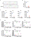
Figure 1.. IFN-κ is upregulated in CLE lesions and lupus keratinocytes.
(A) mRNA expression of type I interferon genes in cutaneous lupus (n=90 CLE, n=13 control). Of the 10 type I family members that had detectable mRNA expression on Affymetrix ST 2.1 array, only IFNK (FDR q<0.001) and IFNA10 (FDR q<0.05) had significantly increased expression in cutaneous lupus (CLE) compared to healthy skin (IFNB was not detectable). (B) Immunofluorescence of CLE lesions revealed IFN-α and IFN-β1 staining in the dermis near the inflammatory infiltrate, whereas IFN-κ expression was seen in the epidermis and dermis of CLE lesions (epidermis is indicated by cytokeratin 14 staining). Representative staining of 3 CLE and 3 controls are shown. (C) Type I IFN response genes, including MX1 and OASL are increased in CLE lesional biopsies (FDR q<0.001) (n=90 CLE, n=13 control). (D) In skin lesions of CLE compared to healthy skin, there is increased phosphorylation of both STAT1 (pSTAT1; red) and STAT2 (pSTAT2; red). Representative staining of 3 CLE and 3 controls are shown. (E) The IFN response protein MX1 was prominently expressed in CLE skin and exhibited co-localization with IFN-κ in the epidermis (as evidenced by yellow color).
Figure 2.
Figure 2.. Keratinocytes exhibit baseline type I interferon activity and express IFN-κ.
(A) Unstimulated normal human keratinocytes (NHK1–3) exhibit heightened expression of type I IFN target genes compared to dermal fibroblasts (FB1–3) and dermal endothelial cells (EC1–4) as measured by RNA-seq (FDR=6.8E-04). (B) RNA-seq reveals IFNK as the only type I IFN detectable in the three skin-derived cell types, and its expression was limited to keratinocytes (C) Immunofluorescent co-localization with the epidermal marker cytokeratin 14 showed co-localization with IFN-κ in healthy epidermis, but no expression of IFN-α, or IFN-β1. Representative of 3 slides is shown (D) The type I IFN response could be transferred to fibroblasts following exposure to keratinocyte conditioned medium for 24hrs (KC-CM), but not non-conditioned keratinocyte medium (KC-M) or conditioned fibroblast medium (FB-CM), as determined by expression of MX1, IRF7, and OASL. This response could be effectively inhibited by addition of neutralizing anti-IFN-κ antibody (10μg/ml), but not with isotype (KC-CM+Iso), anti-IFN-α (KC-CM-IFN-α_Ab) or anti-IFkN-β1(KC-CM-IFNβ1_Ab) antibodies (10μg/ml) (n=3 for all). Data shown with SEM, *p<0.05, **p<0.01, ***p<0.001 (unpaired two-sided Student’s t-test). n=3 for each experiment.
Figure 3.
Figure 3.. IFNK knock-out abolishes type I interferon activity in keratinocytes.
(A) A guide RNA targeting a 20nt sequence in exon 1 of the IFNK gene was designed and knock-out was performed via non-homologous end-joining repair (NHEJ) resulting in the insertion of a single nucleotide (A) in the IFNK gene. (B) Absence of the secreted IFN-κ protein was confirmed by ELISA. (C) IFNK KO suppressed baseline type I IFN activity in unstimulated keratinocytes as determined by expression of MX1, IRF7, IRF9 and OASL. Data shown with SEM, **p<0.01, ***p<0.001, n=3 for all). (D) Baseline type I IFN activity was measured in unstimulated WT and TYK2 KO keratinocytes as determined by expression of IFNK, MX1, IRF7, IRF9 and OASL via real-time PCR. Similar expression changes were measured after treatment with either IFN-α (5 ng/ml) or IFN-κ (5 ng/ml). Data shown with SEM, **p<0.01, ***p<0.001, n=3 for all (unpaired two-sided Student’s t-test). WT: N/TERTs (immortalized human keratinocytes); IFNK KO: IFNK knock-out keratinocytes; TYK2 KO: TYK2 knock-out keratinocytes.
Figure 4.
Figure 4.. IFN-κ is required for rapid keratinocyte responses to exogenous IFN-α.
(A, B) WT, IFNK KO or TYK2 KO KCs were treated with or without IFN-α for the indicated time points followed by Western blot for phosphorylated and total STAT1 and STAT2. Quantification of phosphorylation as compared to WT is shown in (b). (C) WT or IFNK KO KCs were treated with the indicated concentrations of IFN-α followed by qRT-PCR for the indicated genes. Data shown with SEM, *p<0.05, **p<0.01, ***p<0.001, n=3 for all (unpaired two-sided Student’s t-test). WT: N/TERTs (immortalized human keratinocytes); IFNK KO: IFNK knock-out keratinocytes; TYK2 KO: TYK2 knock-out keratinocytes.
Figure 5.
Figure 5.. CLE keratinocytes have heightened basal IFN-κ expression and type I IFN activity.
(A) Cultured keratinocytes from uninvolved skin of patients with SLE and a history of cutaneous lupus exhibited increased protein expression of IFN-κ along with increased pSTAT1 and pSTAT2 activity (representative data from two (out of 3) control and two (out of 3) SLE patients shown). (B) Fibroblasts were treated with KC media alone (KC-M), conditioned media from healthy control KCs (HC-KC-CM) or conditioned media from SLE-KCs (SLE-KC-CM) (n=3 each in triplicate) for 24 hours followed by qRT-PCR for IFN-regulated genes MX1, IRF7, and OASL. (C) KCs stably overexpressing IFN-κ were generated; representative Western Blot of IFN-κ overexpressing line (IFN-κ OE) (both native and GFP-tagged) is shown. (D) MX1 mRNA expression in IFN-κ OE compared to N/TERTs. (E) IFN-κ OE or N/TERTs were grown in the presence or absence of a neutralizing antibody to IFN-κ for 3 days followed by stimulation with 5 ng/mL IFN-α for the indicated time points. MX1 expression was assessed via real-time PCR, normalized to RPLP0 expression and expressed as percent change over unstimulated cells. IFN-K_Ab: IFN-κ antibody; Iso=Isotype control for IFN-κ antibody; Data shown with SEM, *p<0.05, **p<0.01, ***p<0.001, n=3 for all (unpaired two-sided Student’s t-test).
Figure 6.
Figure 6.. IFN-κ promotes UVB induced apoptosis in CLE.
(A) TUNEL staining of normal and CLE skin shows prominent apoptosis in CLE epidermis. Figure is representative of 3 samples. (B) Healthy control and non-lesional SLE KCs (n=6 each) were treated with or without 50mJ/cm2UVB followed by TUNEL staining after 8 hours of culture. Representative photos are shown on the right. (C) IFNK-OE KCs or N/TERTs were treated with UVB followed by TUNEL staining as in (b). (n=5 each). (D) IFNK KO or N/TERT KCs were treated with UVB followed by TUNEL staining as in (b) (n=6 each). (E) Control or SLE KCs (n=5 each) were treated with or without baricitinib (BARI) followed by treatment with 50mJ/cm2UVB followed by TUNEL staining after 8 hours of culture. (F) Condition medium from control or SLE UVB-irradiated KCs was added to monocyte-derived dendritic cell cultures in the presence or absence of baricitinib followed by measurement of CD80 surface expression by flow cytometry and CD80 and MX1 mRNA expression via RT-PCR. Red dots=SLE keratinocyte conditioned media (three different patients), Black dots=control conditioned media (three different healthy controls). All statistical comparisons made via unpaired two-sided Student’s t-test). (G) Immunofluorescent staining denotes increased expression of CD80 on CD11c+ dendritic cells in CLE skin lesions. (H) Schematic outline of the role of IFN-κ in SLE. In control KCs, a basal level of IFN-κ maintains tonic low-level IFN signaling and primes for IFN responses. In SLE KCs, increased autocrine IFN-κ expression drives an elevated tonic IFN response and primes for rapid, robust IFN responses and apoptosis to UVB. Secreted IFN-κ can activate DCs to stimulate immune responses in the skin.

References

    1. Klein R, Moghadam-Kia S, Taylor L, et al. Quality of life in cutaneous lupus erythematosus. J Am Acad Dermatol 2011;64(5):849–58. doi: 10.1016/j.jaad.2010.02.008 [published Online First: 2011/03/15] - DOI - PMC - PubMed
    1. Sanders CJ, Van Weelden H, Kazzaz GA, et al. Photosensitivity in patients with lupus erythematosus: a clinical and photobiological study of 100 patients using a prolonged phototest protocol. Br J Dermatol 2003;149(1):131–7. doi: 10.1046/j.1365-2133.2003.05379.x [pii] [published Online First: 2003/08/02] - DOI - PubMed
    1. Braunstein I, Klein R, Okawa J, et al. The interferon-regulated gene signature is elevated in subacute cutaneous lupus erythematosus and discoid lupus erythematosus and correlates with the cutaneous lupus area and severity index score. British Journal of Dermatology 2012;166(5):971–75. doi: 10.1111/j.1365-2133.2012.10825.x - DOI - PMC - PubMed
    1. Meller S, Winterberg F, Gilliet M, et al. Ultraviolet radiation-induced injury, chemokines, and leukocyte recruitment: An amplification cycle triggering cutaneous lupus erythematosus. Arthritis Rheum 2005;52(5):1504–16. doi: 10.1002/art.21034 - DOI - PubMed
    1. Wenzel J, Tuting T. Identification of type I interferon-associated inflammation in the pathogenesis of cutaneous lupus erythematosus opens up options for novel therapeutic approaches. Exp Dermatol 2007;16(5):454–63. doi: 10.1111/j.1600-0625.2007.00556.x - DOI - PubMed

Publication types

MeSH terms