. 2018 Sep 14;293(37):14237-14248.
doi: 10.1074/jbc.RA118.004174. Epub 2018 Jul 18.

Iron-dependent cleavage of ribosomal RNA during oxidative stress in the yeast Saccharomyces cerevisiae

Affiliations

Iron-dependent cleavage of ribosomal RNA during oxidative stress in the yeast Saccharomyces cerevisiae

Jessica A Zinskie et al. J Biol Chem. .

Abstract

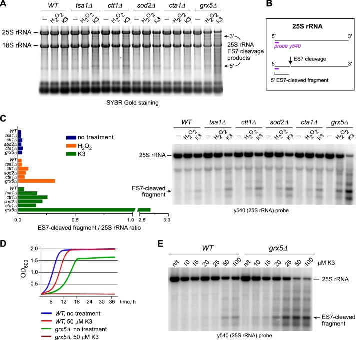
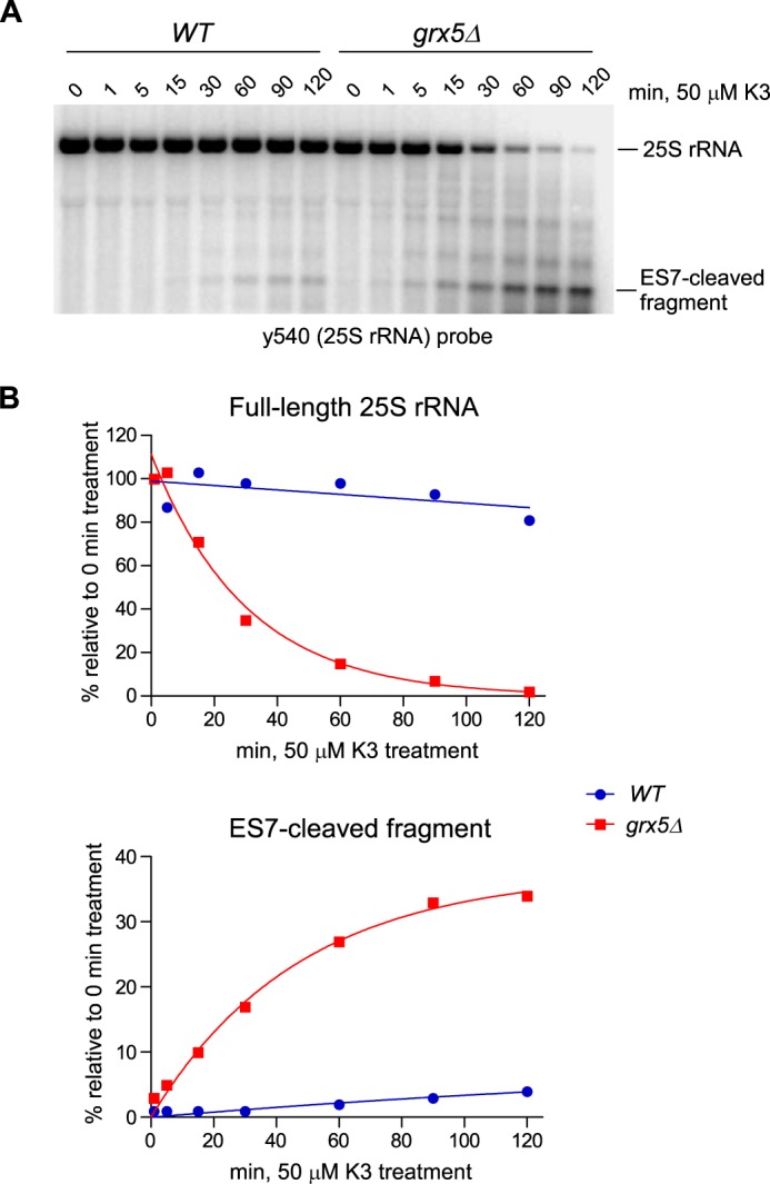
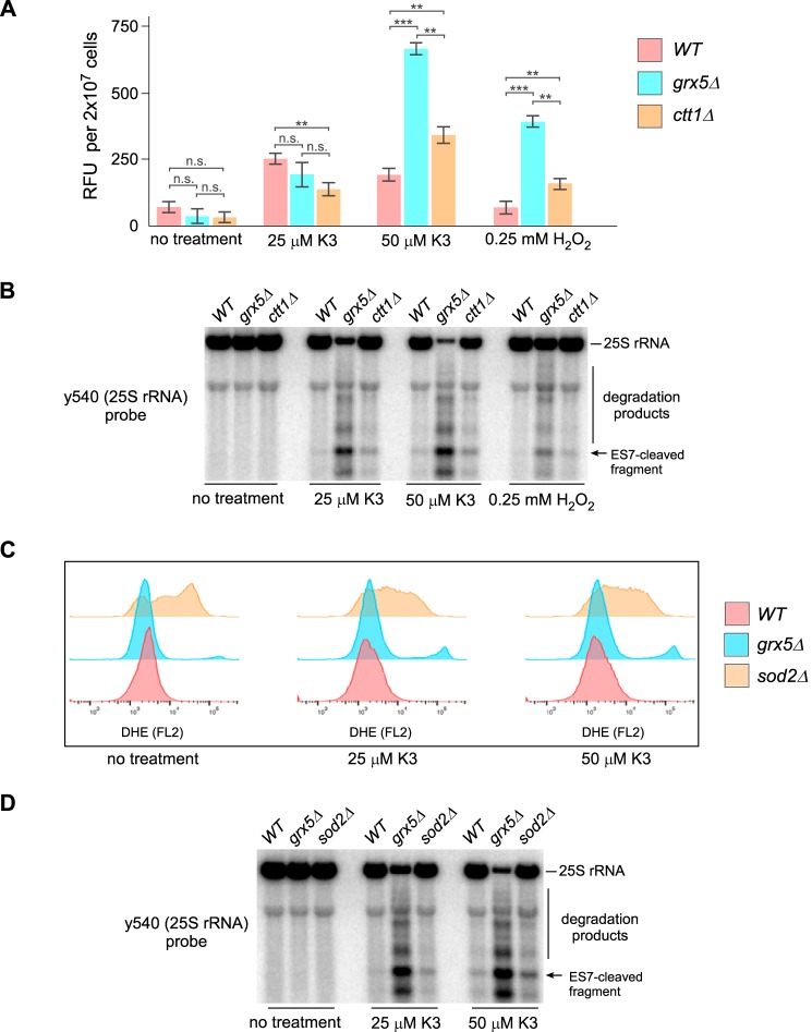
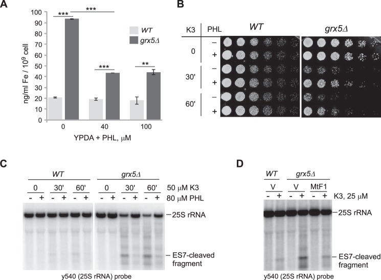
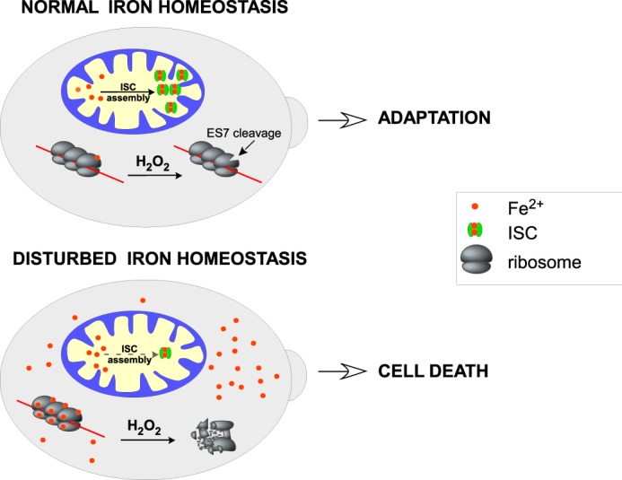
Stress-induced strand breaks in rRNA have been observed in many organisms, but the mechanisms by which they originate are not well-understood. Here we show that a chemical rather than an enzymatic mechanism initiates rRNA cleavages during oxidative stress in yeast (Saccharomyces cerevisiae). We used cells lacking the mitochondrial glutaredoxin Grx5 to demonstrate that oxidant-induced cleavage formation in 25S rRNA correlates with intracellular iron levels. Sequestering free iron by chemical or genetic means decreased the extent of rRNA degradation and relieved the hypersensitivity of grx5Δ cells to the oxidants. Importantly, subjecting purified ribosomes to an in vitro iron/ascorbate reaction precisely recapitulated the 25S rRNA cleavage pattern observed in cells, indicating that redox activity of the ribosome-bound iron is responsible for the strand breaks in the rRNA. In summary, our findings provide evidence that oxidative stress-associated rRNA cleavages can occur through rRNA strand scission by redox-active, ribosome-bound iron that potentially promotes Fenton reaction-induced hydroxyl radical production, implicating intracellular iron as a key determinant of the effects of oxidative stress on ribosomes. We propose that iron binding to specific ribosome elements primes rRNA for cleavages that may play a role in redox-sensitive tuning of the ribosome function in stressed cells.

Keywords: Fenton reaction; RNA; RNA degradation; cell death; glutaredoxin; iron; iron homeostasis; iron metabolism; oxidative stress; reactive oxygen species (ROS); ribosome; yeast.


Conflict of interest statement

The authors declare that they have no conflicts of interest with the contents of this article. The content is solely the responsibility of the authors and does not necessarily represent the official views of the National Institutes of Health

Figures

Figure 1.
Figure 1.
Treatment of grx5Δ cells with oxidants leads to extensive rRNA degradation and growth inhibition. A, increased rRNA degradation in strains deficient for antioxidant genes. Cells in mid-log phase were either left untreated (−) or treated with 50 μm menadione (K3) for 2 h or with 0.25 mm H2O2 for 30 min. Equal amounts of total RNA were separated on an agarose gel and stained with SYBR Gold. B, schematic representation of the sites of ES7 cleavage and probe y540 hybridization in 25S rRNA. C, cells lacking GRX5 demonstrate extensive ES7 cleavage in 25S rRNA. The same RNA samples as shown in A were analyzed by Northern hybridization with the 32P-labeled probe y540. Ratios of the ES7 cleavage 5′ fragment to full-length 25S rRNA (left panel) were obtained by phosphorimaging quantification of the corresponding bands (right panel). D, growth of grx5Δ cells is inhibited by 50 μm menadione. Mid-log cultures were diluted to A600 ∼0.1 and grown at 30 °C with shaking in YPDA in the absence or presence of 50 μm menadione. A600 of the cultures was measured every 5 min for 36 h, and representative growth curves are shown. E, ES7-cleaved fragment formation is detectable in a grx5Δ strain at lower menadione concentrations than in the WT. WT and grx5Δ cells were treated with the indicated concentrations of menadione for 2 h. 25S rRNA was analyzed as in C.
Figure 2.
Figure 2.
25S rRNA fragmentation occurs rapidly in grx5Δ cells in response to oxidant treatment. A, RNA was sampled from mid-log cultures treated with 50 μm menadione (K3) for the indicated times. The full-length 25S rRNA and the ES7-cleaved fragment were detected by Northern hybridization using probe y540. B, change in relative levels of the full-length 25S rRNA and 5′ ES7-cleaved fragment over time. The phosphorimaging signals of the RNA bands in A were fit to a one-phase decay/growth model.
Figure 3.
Figure 3.
Lack of direct correlation between ROS levels and the extent of 25S rRNA fragmentation. A, relative amounts of intracellular H2O2 as determined by an Amplex Red assay. Mid-log WT, grx5Δ, and ctt1Δ cells grown in YPDA were treated with 25 or 50 μm menadione (K3) for 2 h, 0.25 mm H2O2 for 30 min, or remained untreated. Data show mean relative fluorescence units (RFU) in three technical replicates; error bars represent S.D. Two-tailed Student's t test was used for statistical analysis. **, p < 0.01; ***, p < 0.001; n.s., not significant. B, Northern hybridization analysis of 25S rRNA fragmentation from the same cultures as in A. SYBR Gold staining of the gel is shown in Fig. S4A. The experiment shown in A and B was performed twice with similar results. C, analysis of superoxide levels in WT, grx5Δ, and sod2Δ strains treated with menadione as above. Cells were stained with DHE and analyzed by flow cytometry using FlowJo software. D, Northern hybridization analysis of 25S rRNA fragmentation from the same cultures as in C. SYBR Gold staining of the gel is shown in Fig. S4B.
Figure 4.
Figure 4.
Iron overload contributes to oxidant sensitivity and the degradation of ribosomes in grx5Δ cells. A, the intracellular iron level is increased in grx5Δ cells. Overnight cultures were diluted with YPDA to A600 ∼0.2, grown for 4 h, and treated or not with PHL for 1 h. Data show mean values in three biological replicates; error bars represent S.D. Two-tailed Student's t test was used for statistical analysis. **, p < 0.01; ***, p < 0.001. B, PHL restores viability in menadione-treated grx5Δ cells. Cultures grown as in A were treated with 50 μm menadione for 30 or 60 min. Where indicated, cultures were pretreated with 80 μm PHL for 30 min prior to oxidant addition. After treatments, cells were washed, and 2 × 10⁶ cells were serially diluted (1:5) and plated on YPDA agar plates. The plates were incubated at 30 °C for 2 days (WT) or 3 days (grx5Δ). C, PHL pretreatment suppresses the 25S rRNA degradation phenotype. RNA was extracted from the cultures shown in B and analyzed by Northern hybridization with probe y540. SYBR Gold staining of the gel is shown in Fig. S4C. D, suppression of rRNA degradation by ferritin expression. Cells transformed with an empty vector (V) or a mammalian ferritin expression construct (MtF1) were treated or not with 25 μm menadione for 2 h. RNA was analyzed as in C. SYBR Gold staining of the gel is shown in Fig. S4D.
Figure 5.
Figure 5.
25S rRNA undergoes iron-mediated ES7 cleavage in vitro. A, left panel, ribosomal location of the yeast ES7 expansion segment, with surface representation of the a, b, and c arms of 25S rRNA in blue, orange, and red, respectively. ES7CJ is the tripartite junction region. Right panel, enlarged image of the junction, with ES7c in gray and ES7a-b in orange. Nucleotides A611 and U612 at the cleavage site are shown in red and magenta, respectively. Mg2+ ions in the vicinity to the cleavage site are shown as blue spheres. B, left, in vitro cleavage of purified ribosomes with 500, 50, or 5 μm ascorbic acid. Where indicated, reactions contained 1 μm Fe(NH4)2(SO4)2. To chelate iron, ribosomes were pretreated with 1 mm DFO. Reaction products were analyzed by Northern blotting with probe y540. Right, rRNA from grx5Δ cells left untreated or treated with 50 μm menadione or 0.25 mm H2O2 was loaded for comparison on the same gel. C, primer extension analysis of the ES7 region in 25S rRNA after in vitro cleavage with 50 μm ascorbic acid and 1 μm Fe(NH4)2(SO4)2. Control RNAs were extracted from WT cells, grx5Δ cells treated with 50 μm menadione, and tsa1Δ cells treated with 20 mm DTT. Primer extensions were separated on a 6% polyacrylamide/urea gel next to a sequencing ladder. D, ribosomes were treated with 50 or 500 μm ascorbic acid in the presence of 1 μm Fe(NH4)2(SO4)2 in the absence (−) or presence of 100 μm metal chelators EDTA, PHL or BPS. RNA was analyzed as described in B.
Figure 6.
Figure 6.
A working model of the dual role of iron in ribosomes. In cells with balanced iron metabolism, ROS cause limited, position-specific cleavages in rRNA at the sites of bound iron. This might affect ribosome function and optimize translation to conditions of oxidative stress, potentially serving as a stress adaptation mechanism. Accumulation of labile iron, such as in cells with defects in mitochondrial iron metabolism, increases iron binding to ribosomes and leads to extensive rRNA fragmentation, causing global inhibition of translation, which, in turn, may accelerate cell death.

References

    1. D'Autréaux B., and Toledano M. B. (2007) ROS as signalling molecules: mechanisms that generate specificity in ROS homeostasis. Nat. Rev. Mol. Cell Biol. 8, 813–824 10.1038/nrm2256 - DOI - PubMed
    1. Marinho H. S., Real C., Cyrne L., Soares H., and Antunes F. (2014) Hydrogen peroxide sensing, signaling and regulation of transcription factors. Redox Biol. 2, 535–562 10.1016/j.redox.2014.02.006 - DOI - PMC - PubMed
    1. Willi J., Küpfer P., Evéquoz D., Fernandez G., Katz A., Leumann C., and Polacek N. (2018) Oxidative stress damages rRNA inside the ribosome and differentially affects the catalytic center. Nucleic Acids Res. 10.1093/nar/gkx1308 - DOI - PMC - PubMed
    1. Topf U., Suppanz I., Samluk L., Wrobel L., Böser A., Sakowska P., Knapp B., Pietrzyk M. K., Chacinska A., and Warscheid B. (2018) Quantitative proteomics identifies redox switches for global translation modulation by mitochondrially produced reactive oxygen species. Nat. Commun. 9, 324 10.1038/s41467-017-02694-8 - DOI - PMC - PubMed
    1. Ding Q., Markesbery W. R., Cecarini V., and Keller J. N. (2006) Decreased RNA, and increased RNA oxidation, in ribosomes from early Alzheimer's disease. Neurochem. Res. 31, 705–710 10.1007/s11064-006-9071-5 - DOI - PubMed

Publication types

MeSH terms

Associated data