Review
. 2018 Jul 18;145(14):dev156034.
doi: 10.1242/dev.156034.

Polyploidy in tissue homeostasis and regeneration

Affiliations
Review

Polyploidy in tissue homeostasis and regeneration

Jan Inge Øvrebø et al. Development. .

Abstract

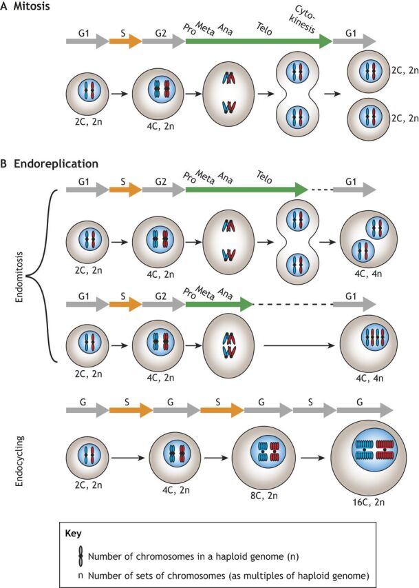
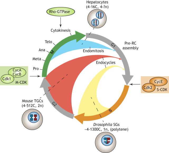
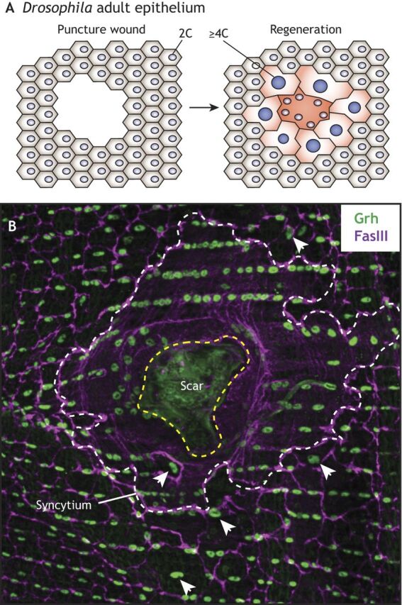
Polyploid cells, which contain multiple copies of the typically diploid genome, are widespread in plants and animals. Polyploidization can be developmentally programmed or stress induced, and arises from either cell-cell fusion or a process known as endoreplication, in which cells replicate their DNA but either fail to complete cytokinesis or to progress through M phase entirely. Polyploidization offers cells several potential fitness benefits, including the ability to increase cell size and biomass production without disrupting cell and tissue structure, and allowing improved cell longevity through higher tolerance to genomic stress and apoptotic signals. Accordingly, recent studies have uncovered crucial roles for polyploidization in compensatory cell growth during tissue regeneration in the heart, liver, epidermis and intestine. Here, we review current knowledge of the molecular pathways that generate polyploidy and discuss how polyploidization is used in tissue repair and regeneration.

Keywords: Endocycling; Endomitosis; Healing; Hippo; Polyploid; Wound.


Conflict of interest statement

Competing interestsThe authors declare no competing or financial interests.

Figures

Fig. 1. An overview of alternative cell cycles. (A,B) While mitosis (A) gives rise to diploid cells, a common path to polyploidy is endoreplication (B), which includes two subgroups: endomitosis and endocycling. Similar to mitotic cells, cells that undergo endomitosis enter the cell cycle, which consists of four canonical phases: G1, S, G2 and M. Endomitosis is characterized by incomplete cytokinesis, thus resulting in a polyploid binucleate cell or a polyploid mononucleate cell. By contrast, endocycling cells lack M phase altogether, resulting in a two-phase cell cycle consisting of alternating G and S phases. Endocycling cells often over- or under-amplify certain genomic regions, resulting in joined polytene chromosomes.
Fig. 1.
An overview of alternative cell cycles. (A,B) While mitosis (A) gives rise to diploid cells, a common path to polyploidy is endoreplication (B), which includes two subgroups: endomitosis and endocycling. Similar to mitotic cells, cells that undergo endomitosis enter the cell cycle, which consists of four canonical phases: G1, S, G2 and M. Endomitosis is characterized by incomplete cytokinesis, thus resulting in a polyploid binucleate cell or a polyploid mononucleate cell. By contrast, endocycling cells lack M phase altogether, resulting in a two-phase cell cycle consisting of alternating G and S phases. Endocycling cells often over- or under-amplify certain genomic regions, resulting in joined polytene chromosomes.
Fig. 2. Alternative cell cycles found in development and regeneration. The mitotic cell cycle is composed of four phases: G1, S, G2 and M. The G1/S and G2/M cell cycle transitions are controlled by S-CDK and M-CDK activities, respectively. Endopolyploidy arises from altered cell cycles in which different cell cycle phases are truncated or bypassed entirely (blue, red and yellow arrows). As examples, mouse hepatocytes skip only cytokinesis in a cell cycle variant known as endomitosis (blue arrow), mouse trophoblast giant cells (TGCs) enter G1 following G2 (red arrow) and Drosophila salivary gland cells (SGs) re-enter a G1-like phase before fully completing DNA replication in S phase (yellow arrow). Endocycles and endomitoses are frequently regulated through downregulation of M-CDK and cytokinesis, respectively.
Fig. 2.
Alternative cell cyclesfound in development and regeneration. The mitotic cell cycle is composed of four phases: G1, S, G2 and M. The G1/S and G2/M cell cycle transitions are controlled by S-CDK and M-CDK activities, respectively. Endopolyploidy arises from altered cell cycles in which different cell cycle phases are truncated or bypassed entirely (blue, red and yellow arrows). As examples, mouse hepatocytes skip only cytokinesis in a cell cycle variant known as endomitosis (blue arrow), mouse trophoblast giant cells (TGCs) enter G1 following G2 (red arrow) and Drosophila salivary gland cells (SGs) re-enter a G1-like phase before fully completing DNA replication in S phase (yellow arrow). Endocycles and endomitoses are frequently regulated through downregulation of M-CDK and cytokinesis, respectively.
Fig. 3. Mitotic-endocycle transitions in Drosophila. Cell growth in Drosophila is controlled by multiple pathways, including the PI3K/TOR, EGFR/MAPK, JAK/STAT, JNK and Hippo (Hpo)/Yki pathways; pathways shown to operate in ovarian follicle cells (O), adult midgut (G), salivary gland (S) and epidermis (E) are indicated. Cellular growth rates affect levels of E2F1 protein, which controls G1/S in a rate-limiting manner through transcriptional activation of Cyclin E (CycE), which binds to and activates CDK2, forming active S-CDK complexes. S phase triggers proteasomal degradation of E2F1 through activation of CRL4cdt2. The G2/M transition is regulated by CycA- and CycB-dependent CDK1 kinase activity (M-CDK), which requires activation by String (Stg; a Cdc25-type phosphatase). M-CDK kinase activity activates the APC/C subunit Fizzy (Fzy). The E3 ligase APC/CFzy targets Geminin (Gem), CycA and CycB for proteasomal degradation; the subsequent depletion of Geminin and M-CDK activity relieves inhibition of Cdt1 and creates a window of low CDK activity (not shown), respectively, which allows re-assembly of the pre-replication complex (pre-RC). In the adult midgut (G) and follicle cells of the ovary (O), mitosis-endocycle transitions are triggered through expression of the Notch ligand Delta in adjacent cells. In follicle cells, active Notch induces Hindsight (Hnt), which represses Stg expression and thereby blocks M-CDK activity. Upregulation of Fizzy-related (Fzr), another activating subunit of APC/C that does not require activation by M-CDK, ensures low M-CDK activity and mediates destruction of Geminin, which in turn allows pre-RC assembly while bypassing M phase. Increased endocycling can also be induced by mechanical stress via the Hippo pathway and JNK. The Hippo pathway stimulates increased ploidy non-cell-autonomously in enterocytes of the adult midgut (G) through expression and secretion of cytokines and growth factors, which activate the EGFR/Ras/MAPK and JAK/STAT pathways, increasing cell growth rates and decreasing the length of G phase. In the adult epidermis (E), Yorkie (Yki) is required for polyploidization cell-autonomously upon wound closure.
Fig. 3.
Mitotic-endocycle transitions in Drosophila. Cell growth in Drosophila is controlled by multiple pathways, including the PI3K/TOR, EGFR/MAPK, JAK/STAT, JNK and Hippo (Hpo)/Yki pathways; pathways shown to operate in ovarian follicle cells (O), adult midgut (G), salivary gland (S) and epidermis (E) are indicated. Cellular growth rates affect levels of E2F1 protein, which controls G1/S in a rate-limiting manner through transcriptional activation of Cyclin E (CycE), which binds to and activates CDK2, forming active S-CDK complexes. S phase triggers proteasomal degradation of E2F1 through activation of CRL4cdt2. The G2/M transition is regulated by CycA- and CycB-dependent CDK1 kinase activity (M-CDK), which requires activation by String (Stg; a Cdc25-type phosphatase). M-CDK kinase activity activates the APC/C subunit Fizzy (Fzy). The E3 ligase APC/CFzy targets Geminin (Gem), CycA and CycB for proteasomal degradation; the subsequent depletion of Geminin and M-CDK activity relieves inhibition of Cdt1 and creates a window of low CDK activity (not shown), respectively, which allows re-assembly of the pre-replication complex (pre-RC). In the adult midgut (G) and follicle cells of the ovary (O), mitosis-endocycle transitions are triggered through expression of the Notch ligand Delta in adjacent cells. In follicle cells, active Notch induces Hindsight (Hnt), which represses Stg expression and thereby blocks M-CDK activity. Upregulation of Fizzy-related (Fzr), another activating subunit of APC/C that does not require activation by M-CDK, ensures low M-CDK activity and mediates destruction of Geminin, which in turn allows pre-RC assembly while bypassing M phase. Increased endocycling can also be induced by mechanical stress via the Hippo pathway and JNK. The Hippo pathway stimulates increased ploidy non-cell-autonomously in enterocytes of the adult midgut (G) through expression and secretion of cytokines and growth factors, which activate the EGFR/Ras/MAPK and JAK/STAT pathways, increasing cell growth rates and decreasing the length of G phase. In the adult epidermis (E), Yorkie (Yki) is required for polyploidization cell-autonomously upon wound closure.
Fig. 4. Mitotic-endocycle transitions in trophoblast giant cells. Cellular growth rates affect activation of E2F1-3, which control G1/S through transcriptional activation of S-CDK activity. E2F1-3 also activate expression of E2F7-8, which in turn repress expression of E2F1 and its targets, thus forming a negative-feedback loop. E2F1-3 are also required for the expression of cyclin A and cyclin B (CcnA and CcnB), which activate CDK1 (M-CDK) and are required for M-phase entry. M-CDK kinase activity activates the APC/C subunit Cdc20. The E3 ligase APC/CCdc20 targets geminin, CcnA and CcnB for proteasomal degradation. Depletion of geminin and M-CDK activity relieves inhibition of Cdt1 and creates a window of low CDK activity, respectively, which allows re-assembly of the pre-replication complex (pre-RC). In vitro, trophoblast stem cells are induced to enter endocycles upon FGF4 deprivation, which reduces CHK1 activity and relieves degradation of p57Kip2 and p21cip1. Accumulation of p57Kip2 and p21cip1 block M-CDK activity, which establish an M-phase bypass and onset of endocycles in TGCs. Further polyploidization is also affected by E2F7-8-dependent downregulation of E2F1-3.
Fig. 4.
Mitotic-endocycle transitions in trophoblast giant cells. Cellular growth rates affect activation of E2F1-3, which control G1/S through transcriptional activation of S-CDK activity. E2F1-3 also activate expression of E2F7-8, which in turn repress expression of E2F1 and its targets, thus forming a negative-feedback loop. E2F1-3 are also required for the expression of cyclin A and cyclin B (CcnA and CcnB), which activate CDK1 (M-CDK) and are required for M-phase entry. M-CDK kinase activity activates the APC/C subunit Cdc20. The E3 ligase APC/CCdc20 targets geminin, CcnA and CcnB for proteasomal degradation. Depletion of geminin and M-CDK activity relieves inhibition of Cdt1 and creates a window of low CDK activity, respectively, which allows re-assembly of the pre-replication complex (pre-RC). In vitro, trophoblast stem cells are induced to enter endocycles upon FGF4 deprivation, which reduces CHK1 activity and relieves degradation of p57Kip2 and p21cip1. Accumulation of p57Kip2 and p21cip1 block M-CDK activity, which establish an M-phase bypass and onset of endocycles in TGCs. Further polyploidization is also affected by E2F7-8-dependent downregulation of E2F1-3.
Fig. 5. Wound-induced polyploidization in the adult Drosophila epithelium. (A) The adult Drosophila epithelium is composed of post-mitotic diploid cells. Upon epithelial puncture wounds, diploid epithelial cells are lost and the open wound is closed by surrounding cells that fuse together to form a syncytium. Cells surrounding the central syncytium undergo endocycles and promote compensatory growth. (B) Immunofluorescence image of regenerating Drosophila adult epithelium. Epidermal nuclei and cell-cell septate junctions, marked by Grainy-head (Grh, in green) and Fas3 (in magenta), respectively, are shown. The boundaries of the scar and syncytium are outlined in yellow and white dashed lines, respectively. Examples of large polyploid nuclei are indicated by arrowheads. Image courtesy of Vicki Losick (see Losick et al., 2013 for details).
Fig. 5.
Wound-induced polyploidization in the adult Drosophila epithelium. (A) The adult Drosophila epithelium is composed of post-mitotic diploid cells. Upon epithelial puncture wounds, diploid epithelial cells are lost and the open wound is closed by surrounding cells that fuse together to form a syncytium. Cells surrounding the central syncytium undergo endocycles and promote compensatory growth. (B) Immunofluorescence image of regenerating Drosophila adult epithelium. Epidermal nuclei and cell-cell septate junctions, marked by Grainy-head (Grh, in green) and Fas3 (in magenta), respectively, are shown. The boundaries of the scar and syncytium are outlined in yellow and white dashed lines, respectively. Examples of large polyploid nuclei are indicated by arrowheads. Image courtesy of Vicki Losick (see Losick et al., 2013 for details).
Fig. 6. Polyploidization during liver regeneration. Hepatocyte growth rates affect the activation of E2F1-3, which controls G1/S through transcriptional activation of cyclin E1 (CcnE1)/Cdk2. E2F1-3 also activate expression of E2F7-8, which in turn repress expression of E2F1 and its targets, thus forming a negative-feedback loop. E2F1-3 are also required for expression of CcnA and CcnB, which activate Cdk1 (M-CDK) and are required for M-phase entry. In hepatocytes, E2F1-3 depletion promotes endoreplication (red arrow), whereas E2F7-8 depletion promotes mitosis (green arrow). Endoreplication is also induced through the Hippo pathway. The Hippo homologs Mst1/2 suppress activity of the Yorkie homologs YAP and TAZ through activation of Lats1/2. Upon Mst1/2 inactivation, YAP promotes Akt activity, which promotes activation of the acetyl transferase p300. The subsequent p300-dependent acetylation of Skp2, an F-box protein of the SCF ubiquitin ligase complex, sequesters Skp2 to the cytoplasm. This prevents proteasomal degradation of p27, an inhibitor of M-CDK activity. Hepatocytes also become polyploid via endomitosis (blue arrow) through downregulation of cytokinetic regulators such as Rho-GTPase. E2F7-8 and miR-122 are known suppressors of cytokinesis and thereby promote endomitosis.
Fig. 6.
Polyploidization during liver regeneration. Hepatocyte growth rates affect the activation of E2F1-3, which controls G1/S through transcriptional activation of cyclin E1 (CcnE1)/Cdk2. E2F1-3 also activate expression of E2F7-8, which in turn repress expression of E2F1 and its targets, thus forming a negative-feedback loop. E2F1-3 are also required for expression of CcnA and CcnB, which activate Cdk1 (M-CDK) and are required for M-phase entry. In hepatocytes, E2F1-3 depletion promotes endoreplication (red arrow), whereas E2F7-8 depletion promotes mitosis (green arrow). Endoreplication is also induced through the Hippo pathway. The Hippo homologs Mst1/2 suppress activity of the Yorkie homologs YAP and TAZ through activation of Lats1/2. Upon Mst1/2 inactivation, YAP promotes Akt activity, which promotes activation of the acetyl transferase p300. The subsequent p300-dependent acetylation of Skp2, an F-box protein of the SCF ubiquitin ligase complex, sequesters Skp2 to the cytoplasm. This prevents proteasomal degradation of p27, an inhibitor of M-CDK activity. Hepatocytes also become polyploid via endomitosis (blue arrow) through downregulation of cytokinetic regulators such as Rho-GTPase. E2F7-8 and miR-122 are known suppressors of cytokinesis and thereby promote endomitosis.

References

    1. Andreassen, P. R., Martineau, S. N. and Margolis, R. L. (1996). Chemical induction of mitotic checkpoint override in mammalian cells results in aneuploidy following a transient tetraploid state. Mutat. Res. 372, 181-194. 10.1016/S0027-5107(96)00138-8 - DOI - PubMed
    1. Anisimov, A. P. (2005). Endopolyploidy as a morphogenetic factor of development. Cell Biol. Int. 29, 993-1004. 10.1016/j.cellbi.2005.10.013 - DOI - PubMed
    1. Aragona, M., Panciera, T., Manfrin, A., Giulitti, S., Michielin, F., Elvassore, N., Dupont, S. and Piccolo, S. (2013). A mechanical checkpoint controls multicellular growth through YAP/TAZ regulation by actin-processing factors. Cell 154, 1047-1059. 10.1016/j.cell.2013.07.042 - DOI - PubMed
    1. Artero, R., Furlong, E. E., Beckett, K., Scott, M. P. and Baylies, M. (2003). Notch and Ras signaling pathway effector genes expressed in fusion competent and founder cells during Drosophila myogenesis. Development 130, 6257-6272. 10.1242/dev.00843 - DOI - PubMed
    1. Audibert, A., Simon, F. and Gho, M. (2005). Cell cycle diversity involves differential regulation of Cyclin E activity in the Drosophila bristle cell lineage. Development 132, 2287-2297. 10.1242/dev.01797 - DOI - PubMed

Publication types

LinkOut - more resources