Review
. 2018 Jul 18;8(3):57.
doi: 10.3390/biom8030057.

Calmodulin: A Multitasking Protein in Kv7.2 Potassium Channel Functions

Affiliations
Review

Calmodulin: A Multitasking Protein in Kv7.2 Potassium Channel Functions

Alessandro Alaimo et al. Biomolecules. .

Abstract

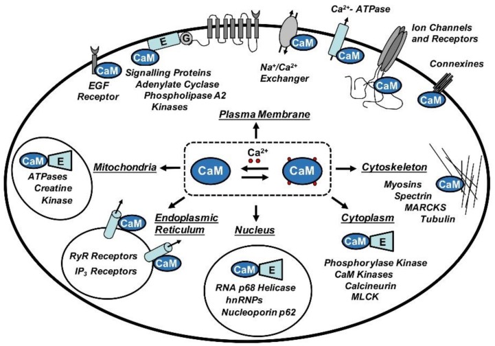
The ubiquitous calcium transducer calmodulin (CaM) plays a pivotal role in many cellular processes, regulating a myriad of structurally different target proteins. Indeed, it is unquestionable that CaM is the most relevant transductor of calcium signals in eukaryotic cells. During the last two decades, different studies have demonstrated that CaM mediates the modulation of several ion channels. Among others, it has been indicated that Kv7.2 channels, one of the members of the voltage gated potassium channel family that plays a critical role in brain excitability, requires CaM binding to regulate the different mechanisms that govern its functions. The purpose of this review is to provide an overview of the most recent advances in structure⁻function studies on the role of CaM regulation of Kv7.2 and the other members of the Kv7 family.

Keywords: Kv7; calcium; calmodulin; potassium channels; signal transduction.


Conflict of interest statement

The authors declare no conflict of interest.

Figures

Figure 1
Figure 1
The central role played by calmodulin (CaM) in the regulation of a wide spectrum of binding partners. Abbreviations: hnRNPs: Heterogeneous nuclear ribonucleoproteins; MARCKS: Myristoylated alanine-rich C-kinase substrate; MLCK: Myosin light-chain kinase.
Figure 2
Figure 2
CaM/Kv7 complexes. Ribbon representation of CaM in complex with AB helices of Kv7 channels. CaM is colored turquoise, the helices A are red, the helices B are blue, the “TW domains” are in dark orange and, finally, Ca2+ and Mg2+ ions are red and magenta, respectively. Structural data were obtained from the Protein Data Bank (PDB), accession codes: (A) CaM/Kv 7.1hAB (4V0C, [66]); (B) CaM/Kv7.3hA-Kv7.2hB (5J03, [69]); (C) CaM/Kv7.1hAB (5VMS, [28]); (D) CaM/Kv7.2hAB (6FEG, [67]); (E) CaM/Kv7.4hAB (6B8L, [59]); (F) CaM/Kv7.5hAB (6B8Q, [59]). Panels A and C were simplified to show only the CaM/hAB complexes. Figures were produced with PyMOL (The PyMOL Molecular Graphics System, Version 1.5.0.4 Schrödinger, LLC.).

References

    1. Cheung W.Y. Cyclic 3′,5′-nucleotide phosphodiesterase. Demonstration of an activator. Biochem. Biophys. Res. Commun. 1970;38:533–538. doi: 10.1016/0006-291X(70)90747-3. - DOI - PubMed
    1. Kakiuchi S., Yamazaki R. Calcium dependent phosphodiesterase activity and its activating factor (PAF) from brain studies on cyclic 3′,5′-nucleotide phosphodiesterase (3) Biochem. Biophys. Res. Commun. 1970;41:1104–1110. doi: 10.1016/0006-291X(70)90199-3. - DOI - PubMed
    1. Jurado L.A., Chockalingam P.S., Jarrett H.W. Apocalmodulin. Physiol. Rev. 1999;79:661–682. doi: 10.1152/physrev.1999.79.3.661. - DOI - PubMed
    1. Chin D., Mens A.R. Calmodulin: A prototypical calcium sensor. Trends Cell Biol. 2000;10:322–328. doi: 10.1016/S0962-8924(00)01800-6. - DOI - PubMed
    1. Saimi Y., Kung C. Calmodulin as an ion channel subunit. Annu. Rev. Physiol. 2002;64:289–311. doi: 10.1146/annurev.physiol.64.100301.111649. - DOI - PubMed

Publication types

LinkOut - more resources