. 2018 Jul 18;8(1):10898.
doi: 10.1038/s41598-018-29170-7.

Quantitative muscle MRI to follow up late onset Pompe patients: a prospective study

Collaborators, Affiliations

Quantitative muscle MRI to follow up late onset Pompe patients: a prospective study

Sebastian Figueroa-Bonaparte et al. Sci Rep. .

Abstract

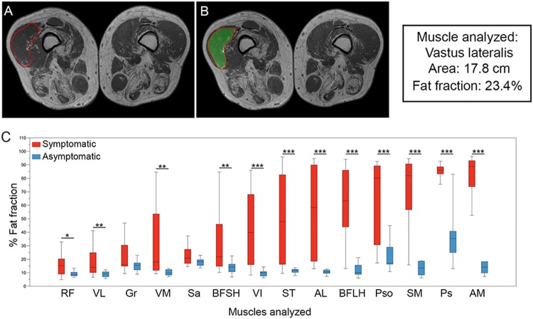
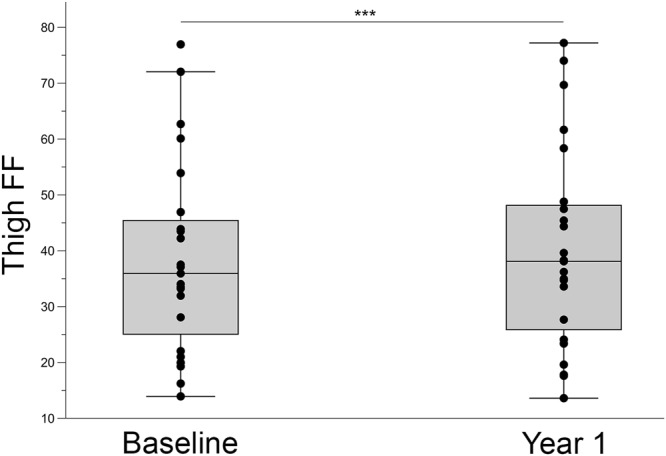
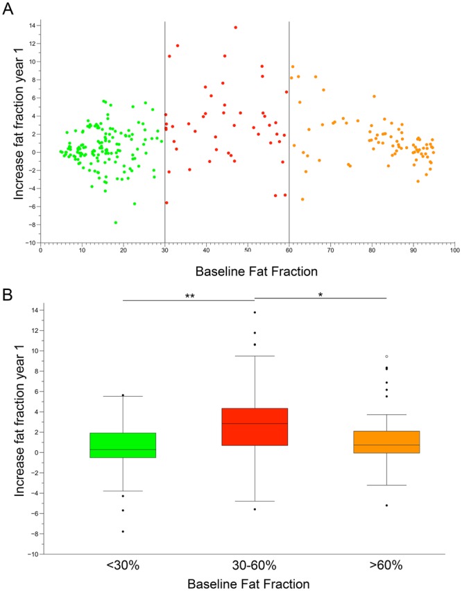
Late onset Pompe disease (LOPD) is a slow, progressive disorder characterized by skeletal and respiratory muscle weakness. Enzyme replacement therapy (ERT) slows down the progression of muscle symptoms. Reliable biomarkers are needed to follow up ERT-treated and asymptomatic LOPD patients in clinical practice. In this study, 32 LOPD patients (22 symptomatic and 10 asymptomatic) underwent muscle MRI using 3-point Dixon and were evaluated at the time of the MRI with several motor function tests and patient-reported outcome measures, and again after one year. Muscle MRI showed a significant increase of 1.7% in the fat content of the thigh muscles in symptomatic LOPD patients. In contrast, there were no noteworthy differences between muscle function tests in the same period of time. We did not observe any significant changes either in muscle MRI or in muscle function tests in asymptomatic patients over the year. We conclude that 3-point Dixon muscle MRI is a useful tool for detecting changes in muscle structure in symptomatic LOPD patients and could become part of the current follow-up protocol in daily clinics.


Conflict of interest statement

The study was supported by Sanofi-Genzyme. The company did not interfere in the design of the protocol or review any data obtained from patients. There are no other conflicts of interest.

Figures

Figure 1
Figure 1
Analysis of fat fraction in thigh and trunk muscles in LOPD patients at baseline. (A,B) Show an example of how ROIs are drawn to obtain total muscle area and fat fraction in 3-point Dixon images. (C) shows the fat fraction calculated for thigh and trunk muscles in symptomatic (red) and asymptomatic (blue) patients. The box plot includes the 25th–75th percentile, the mid lines indicate the median, bars are the 5th–95th percentiles. *P < 0.05, **P < 0.01 and ***P < 0.001. RF: Rectus Femoris, VL: Vastus Lateralis, Gr: Gracilis, VM: Vastus Medialis, Sa: Sartorius, BFSH: Biceps Femoris Short Head, VI: Vastus Intermedius, ST: Semitendinosus, AL: Adductor Longus, BFLH: Biceps Femoris Long Head, Pso: Psoas, SM: Semimembranosus, Ps: Paraspinalis, and AM: Adductor Major.
Figure 2
Figure 2
Yearly progression in thigh muscle fat replacement in symptomatic LOPD patients. The box plot includes the 25th–75th percentile, the mid lines indicate the median, bars are the 5th–95th percentiles. Each dot represents one symptomatic LOPD patient. Mann-Whitney U Test, ***P < 0.001.
Figure 3
Figure 3
Yearly progression of fat replacement in individual muscles of symptomatic patients. (A) Each dot represents fat fraction calculated in a single muscle. Vertical lines divide muscles based on baseline fat fractions: low (green dots, 0–30%), intermediate (red dots, 30–60%) and severe (orange dots, 60–90%). (B) Increase in muscle fat replacement related to baseline fat fraction. The box plot includes the 25th–75th percentile, the mid lines indicate the median, bars correspond to the 5th–95th percentiles. Black dots are outliers. Kruskal-Wallis test, *P < 0.05, **P < 0.01.

References

    1. Bembi B, et al. Diagnosis of glycogenosis type II. Neurology. 2008;71:S4–11. doi: 10.1212/WNL.0b013e31818da91e. - DOI - PubMed
    1. Schuller A, Wenninger S, Strigl-Pill N, Schoser B. Toward deconstructing the phenotype of late-onset Pompe disease. Am J Med Genet C Semin Med Genet. 2012;160C:80–88. doi: 10.1002/ajmg.c.31322. - DOI - PubMed
    1. van der Ploeg AT, et al. A randomized study of alglucosidase alfa in late-onset Pompe’s disease. N Engl J Med. 2010;362:1396–1406. doi: 10.1056/NEJMoa0909859. - DOI - PubMed
    1. van der Ploeg AT, et al. Open-label extension study following the Late-Onset Treatment Study (LOTS) of alglucosidase alfa. Mol Genet Metab. 2012;107:456–461. doi: 10.1016/j.ymgme.2012.09.015. - DOI - PubMed
    1. Gungor D, et al. Impact of enzyme replacement therapy on survival in adults with Pompe disease: results from a prospective international observational study. Orphanet J Rare Dis. 2013;8:49. doi: 10.1186/1750-1172-8-49. - DOI - PMC - PubMed

Publication types