. 2018 Jul 18;9(1):2809.
doi: 10.1038/s41467-018-05253-x.

The solute carrier SLC9C1 is a Na+/H+-exchanger gated by an S4-type voltage-sensor and cyclic-nucleotide binding

Affiliations

The solute carrier SLC9C1 is a Na+/H+-exchanger gated by an S4-type voltage-sensor and cyclic-nucleotide binding

F Windler et al. Nat Commun. .

Erratum in

Abstract

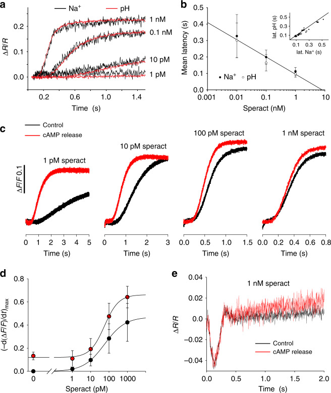
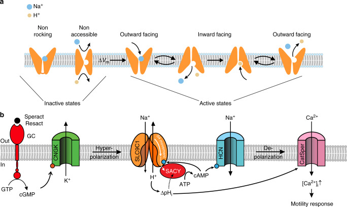
Voltage-sensing (VSD) and cyclic nucleotide-binding domains (CNBD) gate ion channels for rapid electrical signaling. By contrast, solute carriers (SLCs) that passively redistribute substrates are gated by their substrates themselves. Here, we study the orphan sperm-specific solute carriers SLC9C1 that feature a unique tripartite structure: an exchanger domain, a VSD, and a CNBD. Voltage-clamp fluorimetry shows that SLC9C1 is a genuine Na+/H+ exchanger gated by voltage. The cellular messenger cAMP shifts the voltage range of activation. Mutations in the transport domain, the VSD, or the CNBD strongly affect Na+/H+ exchange, voltage gating, or cAMP sensitivity, respectively. Our results establish SLC9C1 as a phylogenetic chimaera that combines the ion-exchange mechanism of solute carriers with the gating mechanism of ion channels. Classic SLCs slowly readjust changes in the intra- and extracellular milieu, whereas voltage gating endows the Na+/H+ exchanger with the ability to produce a rapid pH response that enables downstream signaling events.


Conflict of interest statement

The authors declare no competing interests.

Figures

Fig. 1
Fig. 1
Structural features of SpSLC9C1. a Transmembrane topology of Escherichia coli NhaA (EcNhaA), Methanocaldococcus jannaschii NhaP1 (MjNhaP1), and sea urchin Strongylocentrotus purpuratus SLC9C1 (SpSLC9C1); VSD voltage-sensing domain, CNBD cyclic nucleotide-binding domain. Amino acids that may participate in Na+ coordination are highlighted (black dots). Asterisks indicate conserved Arg residues that are relevant for Na+/H+ antiport; Arg320 and Arg347 in MjNhaP1; Arg399 and Arg431 in SpSLC9C1. An Arg is substituted by Lys300 in EcNhaA. b Scheme of the trigonal bi-pyramidal structure of the Na+ coordination site from MjNhaP1. Numbers refer to the respective amino-acid residues in E. coli (blue), M. jannaschii (red), and S. purpuratus (black). c Sequence comparison of the VSD from several SLC9C1 members with the canonical VSD of Drosophila Shaker K+ channel (DmShaker) and Ciona intestinalis voltage-sensor-containing-phosphatase (CiVSP). S. purpuratus (SpSLC9C1), H. sapiens (HsSLC9C1), M. musculus (MmSLC9C1), C. intestinalis (CiSLC9C1), and spotted gar L. oculatus (LoSLC9C1). Voltage sensors carry conserved positively charged residues in S4 (blue) and conserved negatively charged amino acids in S1–S3 (red). d Cyclic nucleotide-binding domains from sea urchin (SpSLC9C1), human (HsSLC9C1) and mouse (MmSLC9C1) SLC9C1, mouse HCN channel MmHCN2, bovine CNG channel bCNGA1, and C. elegans CNG channel (CeTAX4). The CNBD comprises three α-helices (αA, αB, and αC), eight β-strands (β1–β8), and a phosphate-binding cassette (PBC). Highlighted key residues are the purine-binding residues Val and Leu (β4 and β5), the ribofuranose-binding residues Gly/Glu (β6), the phosphate-binding residues Arg/Thr, and the purine-binding Arg in αC of MmHCN2
Fig. 2
Fig. 2
Gating currents of SpSLC9C1. Voltage protocol, gating currents, and charge–voltage (Q/V) relation. The solid curve in the Q/V relations (insets) represents a Boltzmann fit. a Upper: wt SpSLC9C1 without cNMP (V1/2 = −91.4 mV, slope s= 9.2 mV). Middle: non-transfected CHO cells. Lower: wt SpSLC9C1, 1 mM cAMP in the pipette solution (V1/2 = −75.4 mV, s= 8.2 mV). b Q/Qmax vs. Vm relation. Mean ± SD (n = number of experiments) of V1/2 and slope s was determined by a Boltzmann fit (w/o cNMP: −94.7 ± 2.9 mV, s = 8.5 ± 0.8 mV, qg = 3.1 e0, n = 6; cAMP: −74.4 ± 6.4 mV, s = 8.8 ± 1.9 mV, qg = 2.9 e0, n = 7; cGMP: −86.9 ± 3.0 mV, s = 8.3 ± 1.0 mV, qg = 3.1 e0, n = 7). c Replacing Arg803 in S4 by Gln (R803Q) shifted V1/2 by −24 mV and s by 5 mV (V1/2 = −117.9 ± 7.1 mV, s = 13.0 ± 1.1 mV, qg = 2.0 e0, n = 7). This mutation did not affect V1/2 modulation by 1 mM cAMP (V1/2 = −96.8 ± 6.6 mV, s = 13.5 ± 2.6 mV, qg = 1.9 e0, n = 6). Gray line: voltage dependence of wt SpSLC9C1 without cNMP. d Replacing Arg1053 in the CNBD by Gln (R1053Q) does not affect V1/2 without cNMP (−93.4 ± 1.7 mV, s = 9.4 ± 2.0 mV, qg = 2.8 e0, n = 6), but strongly reduced V1/2 shift by cAMP (V1/2 = −93.9 ± 4.2 mV, s = 10.3 ± 1.0 mV, qg = 2.5 e0, n = 5). Gray line: voltage dependence of wt SpSLC9C1 with cNMP. e In the NHE-domain mutant (R399A), Na+/H+ exchange is abolished (Fig. 5), but not gating currents (Supplementary Figure 4). V1/2 in the absence (−87.3 ± 2.8 mV, s = 10.4 ± 1.8 mV, qg = 2.5 e0, n = 3) and presence of cAMP (−76.5 ± 9.6 mV, s = 6.0 ± 0.8 mV, qg = 4.3 e0, n = 5). Gray line: voltage dependence of wt SpSLC9C1 without cNMP
Fig. 3
Fig. 3
Voltage-clamp pHi fluorimetry of SpSLC9C1 activity. a Alkalinization induced by a 20 s step hyperpolarization to −100 mV using an inwardly directed Na+ gradient ([Na+]i 14 mM; [Na+]o 140 mM; pHi = 7.2; pHo = 7.4, forward mode, red). No pHi change occurred in non-transfected CHO cells (black). b Acidification induced by a 20 s step hyperpolarization to −100 mV using an outwardly directed Na+ gradient ([Na+]i 14 mM; [Na+]o 0 mM; pHi = 7.2; pHo = 7.4, reverse-mode, blue). c Perfusion with symmetric solutions (black line) abolished the net Na+/H+ exchange due to a lacking gradient (pHi = pHo = 7.2, [Na]i = [Na]o = 14 mM). Voltage induced net Na+/H+ exchange was restored when the cell was perfused with 140 mM Na+ (pHo = 7.4). d When Arg399 in T12 of the exchanger domain is replaced by Ala (R399A), Na+/H+ exchange was abolished. e pHi responses to repetitive voltage steps from −40 to −100 mV in a CHO-SpSLC9C1 cell. Dotted line indicates resting pHi. ([Na+]i 14 mM; [Na+]o 140 mM; pHi = 7.2; pHo = 7.4). f Repetitive stimulation of SpSLC9C1 activity (reverse mode) in CHO cells that co-express the H+-selective channel Hv1. Cells quickly recovered from acidification by activation of Hv1 at +47 mV. ([Na+]i 14 mM; [Na+]o 0 mM; pHi = 7.2; pHo = 7.4). g Voltage dependence of SpSLC9C1 activation was determined by stepping Vm between −23 and −103 mV to +47 mV (V1/2 = −70.4 mV; [Na+]i 14 mM; [Na+]o 0 mM; pHi = 7.2; pHo = 7.4). h Normalized ΔR values were plotted against Vm to yield the V1/2 values by a fit with the Boltzmann equation (V1/2 = −70.9 ± 2.5 mV, s = 3.3 ± 0.9 mV, n = 7). Mean values are summarized in Table 1
Fig. 4
Fig. 4
Modulation of SpSLC9C1 activity by cAMP. a Action of 1 mM cAMP in the pipette solution on the voltage dependence of SpSLC9C1 (V1/2 = −53.4 mV). ([Na+]o 0 mM; [Na+]i 14 mM; pHi = 7.2; pHo = 7.4). b Normalized ΔR values were plotted against Vm to yield the V1/2 values by a fit with the Boltzmann equation (w/o cNMP: V1/2 = −70.9 ± 2.5 mV, s = 3.3 ± 0.9 mV, n = 7; cAMP: V1/2 = −56.8 ± 2.7 mV, s= 4.6 ± 1.5 mV, n = 7; cGMP: V1/2 = −67.8 ± 5.4 mV, s= 2.8 ± 1.0 mV, n = 9). c Flash photolysis of caged cAMP enhanced SpSLC9C1 activity at a holding voltage of −63 mV and saturated after 3–4 flashes, same conditions as in a. d Normalized ΔR values were plotted against the number of flashes. Black circles show values from c. Decreasing the light energy of the flash, saturation of SpSLC9C1 activity required 5–6 flashes (white circles). e The shift of V1/2 and s values evoked by flash photolysis were similar to those using cAMP in the pipette (black triangles: V1/2 = −55.3 ± 5.1 mV, s= 5.0 ± 0.6 mV, n = 3; for comparison: w/o cNMP (black circles) and 1 mM cAMP (white circles). f Replacing the Arg1053 in the CNBD by Gln (R1053Q) maintained a wild-type-like V1/2 in the absence of cNMP (−70.8 ± 4.8 mV, s = 3.6 ± 1.1 mV, n = 5), but strongly reduced the V1/2 shift by cAMP (−69.5 ± 3.8 mV, s = 3.9 ± 1.2 mV, n = 6). Mean values are summarized in Table 1
Fig. 5
Fig. 5
Sodium and proton fluxes in S. pupuratus sperm. a Speract-induced changes in fluorescence ratio (ΔR/R) indicating Na+ influx (ANG2, black) or proton efflux (pHi) (BCECF, red); signals were scaled and superimposed. Speract concentrations are shown on the right. b Mean latency ± SD (n = 7) of pHi and Na+ signal plotted vs. speract concentration. Inset: latency of pHi and Na+ signal at different speract concentrations (10 pM black circles; 100 pM white circles; and 1 nM triangles). The line represents the identity function (n = 7). c Speract-induced alkalinization monitored by pHrodo Red fluorescence in the absence (black trace) or presence (red trace) of release of cAMP from DEACM-caged cAMP with a continuously pulsing UV-LED. UV light was applied during the entire recording time. d The maximal slope of the pHrodo Red time course was plotted vs. speract concentration in the absence (black) and presence (red) of cAMP released from DEACM-caged cAMP (n = 3). e Voltage recordings during speract stimulation (1 nM) in the absence (black) or presence (red) of cAMP released from DEACM-caged cAMP
Fig. 6
Fig. 6
Analysis of SpSLC9C1 expression in sea urchin sperm. a Representative western blot using protein lysates from flagella and heads of S. purpuratus sperm, from CHO cells heterologously expressing HA-tagged SpSLC9C1 (SpSLC9C1-HA), and from non-transfected CHO cells (mock). SpSLC9C1 in flagella and heads was probed with the anti-SpSLC9C1 antibody SU2 (left panel) and SpSLC9C1-HA in CHO K1 cells was probed with an HA antibody (right panel, see also Supplementary Figure 8). b Immunocytochemical analysis of sperm stained with SU2 (left) and pre-immune serum control (right). Upper panels: bright-field (DIC) microscopy. Lower panels: overlay of DIC and fluorescence images (SpSLC9C1, green; nucleus stained with DAPI, blue). Scale bars represent 10 µm
Fig. 7
Fig. 7
Models of SLC9C1 gating by voltage and cAMP. a Cartoon depicting that SpSLC9C1 is active only when the voltage enables its activity. The VSD could gate exchange activity by providing access for ions to their binding sites either by removing a physical gate or by enhancing the binding affinity for uploading (non-accessible). Alternatively, in the resting state, ions have access to their binding sites, but the rocking mechanism is locked (non-rocking); voltage unlocks the rocking motion and thus allows switching between outward- and inward facing conformations. b Chemotactic signaling pathway in sea urchin sperm. Chemoattractant binding results in cGMP production and CNGK channel opening. The subsequent hyperpolarization activates SLC9C1. Alkalinization and cAMP production cooperate to open the CatSper channel. The intimate interaction between the exchanger and the soluble adenylate cyclase SACY is illustrated

References

    1. Cesar-Razquin A, et al. A call for systematic research on solute carriers. Cell. 2015;162:478–487. - PubMed
    1. Wang D, King SM, Quill TA, Doolittle LK, Garbers DL. A new sperm-specific Na+/H+ exchanger required for sperm motility and fertility. Nat. Cell Biol. 2003;5:1117–1122. - PubMed
    1. Zhang X, Zeng X, Xia XM, Lingle CJ. pH-regulated Slo3 K+ channels: properties of unitary currents. J. Gen. Physiol. 2006;128:301–315. - PMC - PubMed
    1. Zeng XH, Yang C, Kim ST, Lingle CJ, Xia XM. Deletion of the Slo3 gene abolishes alkalization-activated K+ current in mouse spermatozoa. Proc. Natl Acad. Sci. USA. 2011;108:5879–5884. - PMC - PubMed
    1. Fechner S, et al. A K+-selective CNG channel orchestrates Ca2+ signalling in zebrafish sperm. eLife. 2015;4:e07624. - PMC - PubMed

Publication types

MeSH terms

LinkOut - more resources