. 2018 Jul 4:9:1483.
doi: 10.3389/fmicb.2018.01483. eCollection 2018.

Expression and (Lacking) Internalization of the Cell Surface Receptors of Clostridioides difficile Toxin B

Affiliations

Expression and (Lacking) Internalization of the Cell Surface Receptors of Clostridioides difficile Toxin B

Dennis Schöttelndreier et al. Front Microbiol. .

Abstract

Toxin-producing strains of Clostridioides difficile and Clostridium perfringens cause infections of the gastrointestinal tract in humans and ruminants, with the toxins being major virulence factors, essential for the infection, and responsible for the onset of severe symptoms. C. difficile toxin A (TcdA) and toxin B (TcdB), and the large cytotoxin (TpeL) from C. perfringens are single chain bacterial protein toxins with an AB-like toxin structure. The C-terminal delivery domain mediates cell entry of the N-terminal glycosyltransferase domain by receptor-mediated endocytosis. Several cell surface proteins have been proposed to serve as toxin receptors, including chondroitin-sulfate proteoglycan 4 (CSPG4), poliovirus receptor-like 3 (PVRL3), and frizzled-1/2/7 (FZD1/2/7) for TcdB and LDL-receptor-related protein-1 (LRP1) for TpeL. The expression of the TcdB receptors was investigated in human intestinal organoids (HIOs) and in cultured cell lines. HIOs from four human donors exhibited a comparable profile of receptor expression, with PVRL3, LRP1, and FZD7 being expressed and CSPG4 and FZD2 not being expressed. In human epithelial Caco-2 cells and HT29 cells as well as in immortalized murine fibroblasts, either receptor FZD2/7, CSPG4, PVRL3, and LRP1 was expressed. The question whether the toxins take advantage of the normal turnover of their receptors (i.e., constitutive endocytosis and recycling) from the cell surface or whether the toxins activity induce the internalization of their receptors has not yet been addressed. For the analysis of receptor internalization, temperature-induced uptake of biotinylated toxin receptors into immortalized mouse embryonic fibroblasts (MEFs) and Caco-2 cells was exploited. Solely LRP1 exhibited constitutive endocytosis from the plasma membrane to the endosome, which might be abused by TpeL (and possibly TcdB as well) for cell entry. Furthermore, internalization of CSPG4, PVRL3, FZD2, and FZD7 was observed neither in MEFs nor in Caco-2 cells. FZD2/7, CSPG4, and PVRL3 did thus exhibit no constitutive recycling. The presence of TcdB and the p38 activation induced by anisomycin were not able to induce or enhance CSPG4 or PVRL3 uptake in MEFs. In conclusion, FZD2/7, CSPG4, and PVRL3 seem to serve as cell surface binding receptors rather than internalizing receptors of TcdB.

Keywords: Clostridium perfringens; cell surface; clostridial glycosylating toxins; clostridioides difficile infection; endocytosis; fibroblasts/myofibroblast; human intestinal organoids; receptors.


Figures

FIGURE 1
FIGURE 1
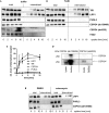
Expression of toxin receptors in human intestinal organoids (HIO) and cell lines. Human intestinal organoids from four donors (D1–4) cultured in matrigel and subconfluent human intestinal epithelial (Caco-2) cells, human colon adenocarcinoma (HT29) cells, and SV40-immortalized mouse embryonic fibroblasts (MEFs) were lysed in Laemmli sample buffer. The expression of the indicated toxin receptors was analyzed by immunoblotting with gp96 serving as loading control. Representative immune-blots are from one of three independent experiments.
FIGURE 2
FIGURE 2
Internalization of toxin receptors. Internalization of reversibly biotinylated cell surface proteins into serum-cultured murine fibroblasts (MEFs) either left non-treated (A) or treated with TcdB (B) was induced by temperature shift to 37°C. Cells were collected at the indicated times. Cells were either left non-treated or exposed to GSH on ice to strip off biotin from still surface-exposed molecules. Biotinylated proteins were retrieved on neutravidin-agarose, eluted, and analyzed by immunoblotting. (C) Internalization of toxin receptors into non-treated fibroblasts was quantified by densitometry of immunoblot (n = 3). (D) Lysate from non-treated fibroblasts (lysate) and immunoprecipitate biotinylated cell surface proteins were subjected to immunoblotting and analyzed with the indicated CSPG4 antibodies. (E) Internalization of biotinylated cell surface proteins was further analyzed in serum-starved fibroblasts pretreated anisomycin (30 μM, E). Representative immunoblots are from one of three independent experiments.
FIGURE 3
FIGURE 3
Internalization of toxin receptors into Caco-2 cells. (A) Internalization of reversibly biotinylated cell surface proteins into serum-cultured Caco-2 cells was induced by temperature shift to 37°C. Cells were collected at the indicated times. Cells were either left non-treated or exposed to GSH on ice to strip off biotin from still surface-exposed molecules. Biotinylated proteins were retrieved on neutravidin-agarose, eluted, and analyzed by immunoblotting. Representative immunoblots are from one of two independent experiments. (B) Internalization of toxin receptors was quantified by densitometry of immunoblot (n = 2).
FIGURE 4
FIGURE 4
Two-receptor-model of TcdB. (1) TcdB binds to (non-internalizing) binding receptors at the cell surface. (Non-internalizing) binding receptors of TcdB are CSPG4, FZD2/7, and PVRL3. (2) The binding receptors form heterodimers with the internalizing receptors. TcdB then also binds to the internalizing receptors. The only candidate for an internalizing TcdB receptor is LRP1. (3) The toxin is released from the binding receptor, allowing the internalization of the TcdB-receptor complex into the early endosome. (4) Upon acidification, the glucosyltransferase domain (GTD) is released through a pore into the cytosol. The GTD mono-O-glucosylates (and thereby inactivates) Rho-GTPases.

References

    1. Aktories K., Schwan C., Jank T. (2017). Clostridium difficile toxin biology. Annu. Rev. Microbiol. 71 281–307. 10.1146/annurev-micro-090816-093458 - DOI - PubMed
    1. Bobo L. D., El Feghaly R. E., Chen Y. S., Dubberke E. R., Han Z., Baker A. H., et al. (2013). MAPK-activated protein kinase 2 contributes to Clostridium difficile-associated inflammation. Infect. Immun. 81 713–722. 10.1128/IAI.00186-12 - DOI - PMC - PubMed
    1. Chandrasekaran R., Lacy D. B. (2017). The role of toxins in Clostridium difficile infection. FEMS Microbiol. Rev. 41 723–750. 10.1093/femsre/fux048 - DOI - PMC - PubMed
    1. Chen P., Tao L., Wang T., Zhang J., He A., Lam K. H., et al. (2018). Structural basis for recognition of frizzled proteins by Clostridium difficile toxin B. Science 360 664–669. 10.1126/science.aar1999 - DOI - PMC - PubMed
    1. Cuadrado A., Nebreda A. R. (2010). Mechanisms and functions of p38 MAPK signalling. Biochem. J. 429 403–417. 10.1042/BJ20100323 - DOI - PubMed