

Ultrastructures of silver nanoparticles biosynthesized using endophytic fungi
- PMID: 30023179
- PMCID: PMC6014216
- DOI: 10.1016/j.jmau.2014.10.004
Ultrastructures of silver nanoparticles biosynthesized using endophytic fungi
Abstract
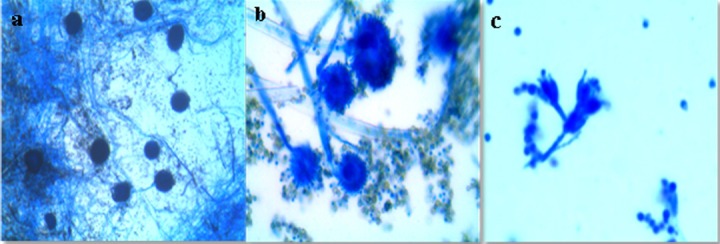
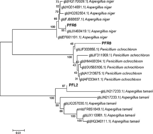
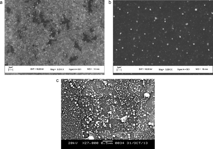
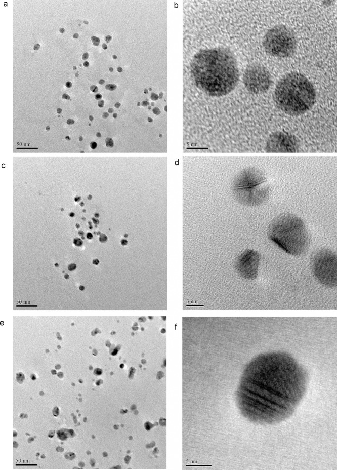
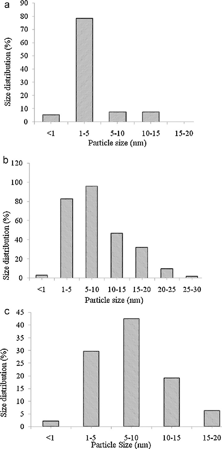
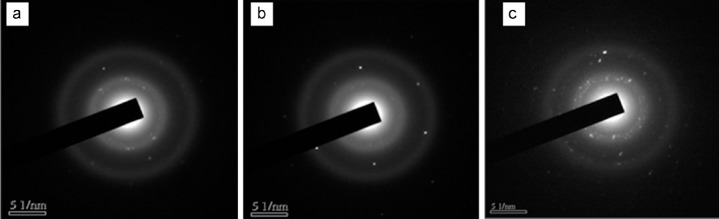
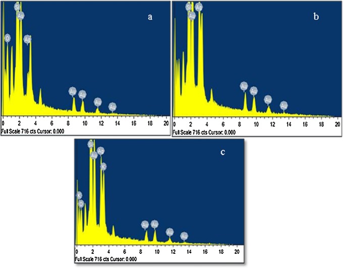
Three endophytic fungi Aspergillus tamarii PFL2, Aspergillus niger PFR6 and Penicllium ochrochloron PFR8 isolated from an ethno-medicinal plant Potentilla fulgens L. were used for the biosynthesis of silver nanoparticles. Scanning and transmission electron microscopic analysis were performed to study the structural morphology of the biosynthesized silver nanoparticles. The electron microscopy study revealed the formation of spherical nanosized silver particles with different sizes. The nanoparticles synthesized using the fungus A. tamarii PFL2 was found to have the smallest average particle size (3.5 ±3 nm) as compared to the nanoparticles biosynthesized using other two fungi A. niger PFR6 and P. ochrochloron PFR8 which produced average particle sizes of 8.7 ±6 nm and 7.7 ±4.3 nm, respectively. The energy dispersive X-ray spectroscopy (EDS) technique in conjunction with scanning electron microscopy was used for the elemental analysis of the nanoparticles. The selected area diffraction pattern recorded from single particle in the aggregates of nanoparticles revealed that the silver particles are crystalline in nature.
Keywords: Crystalline; Electron microscopy; Endophytic fungi; Silver nanoparticles.
Figures
References
-
- Liu J, Aruguete DM, Murayama M, Hochella MF. Influence of size and aggregation on the reactivity of an environmentally and industrially relevant nanomaterial (PbS) Environ Sci Technol. 2009;43:8178–83. - PubMed
-
- Kittler S, Greulich C, Diendorf J, Koller M, Epple M. Toxicity of silver nanoparticles increases during storage because of slow dissolution under release of silver ions. Chem Mater. 2010;22:4548–54.
-
- Zhang W, Yao Y, Sullivan N, Chen YS. Modeling the primary size effects of citrate-coated silver nanoparticles on their ion release kinetics. Environ Sci Technol. 2011;45:4422–8. - PubMed
-
- Mansur HS, Grieser F, Marychurch MS, Biggs S, Urquhart RS, Furlong DN. Photoelectrochemical properties of ‘Q-state’ CdS particles in arachidic acid Langmuir–Blodgett films. J Chem Soc Faraday Trans. 1995;91:665–72.
-
- Chan WCW, NieS Quantum dot bioconjugates for ultrasensitive non-isotopic detection. Science. 1998;281:2016–8. - PubMed
LinkOut - more resources
Full Text Sources
Other Literature Sources