Review
. 2018 Jun;6(11):210.
doi: 10.21037/atm.2018.05.25.

Surgery for intrathoracic tracheoesophageal and bronchoesophageal fistula

Affiliations
Review

Surgery for intrathoracic tracheoesophageal and bronchoesophageal fistula

Benoit Jacques Bibas et al. Ann Transl Med. 2018 Jun.

Abstract

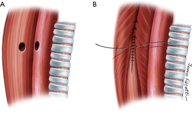
Benign tracheoesophageal fistula (TEF) results from an abnormal communication between the posterior wall of the trachea or bronchi and the adjacent anterior wall of the esophagus. It can be acquired or congenital. The onset of the TEF has a negative impact on the patient's health status and quality of life because of swallowing difficulties, recurrent aspiration pneumonia, and severe weight loss. Several acquired conditions may cause TEF. The most frequent is prolonged orotracheal intubation (75% of the cases). Usually, there is an erosion of the tracheal and esophageal wall by the continuous pressure between the endotracheal tube and the esophageal wall; particularly in the presence of a nasogastric or feeding tube within the esophageal lumen. Furthermore, tracheal stenosis is often associated, and adds complexity to the disease. Preparation for the surgical procedure may take weeks or even months. It includes definitive weaning from mechanical ventilation, treatment of respiratory infection, physiotherapy, and correction of malnutrition through enteral feeding. Surgical repair of a TEF is an elective procedure. It consists of division of the fistula, suture of the esophagus and trachea and protection of the suture lines with a buttressed muscle flap. TEF repair is a complex and challenging procedure, thus, high morbidity and mortality are expected. Nonetheless, surgical management yields excellent long-term results, and it should be considered the first-line treatment for this condition. Definitive fistula closure occurs in about 90-95% of the cases.

Keywords: Trachea; tracheal stenosis; tracheoesophageal fistula (TEF).


Conflict of interest statement

Conflicts of Interest: The authors have no conflicts of interest to declare.

Figures

Figure 1
Figure 1
Gastrobronchial fistula. (A) Post-esophagectomy fistula from the gastric conduit to the right main bronchus; (B) bronchoscopy image; (C) failed attempted endoscopic closure of the fistula with a self-expanding metallic stent; (D) CT scan shows signs of pulmonary soilage and aspiration pneumonia
Figure 2
Figure 2
CT scan—sagittal reconstruction. (A) Tracheoesophageal fistula (arrow) with tracheostomy; (B) a silicone T-tube is used to block the fistula and restore nasal airflow.
Figure 3
Figure 3
A cervicotomy is usually sufficient to repair most TEFs. The fistula site (or tracheostomy) can be included in the incision, as shown. A partial sternotomy can be performed, if necessary. TEF, tracheoesophageal fistula.
Figure 4
Figure 4
Types of tracheoesophageal fistula. (A) Large intrathoracic TEF (10 mm) without tracheal stenosis. A partial sternotomy was necessary for the surgical repair; (B) TEF located in the cervicothoracic transition, with tracheal stenosis; (C) large cervicothoracic TEF without tracheal stenosis. TEF, tracheoesophageal fistula.
Figure 5
Figure 5
Lateral approach to a tracheoesophageal fistula. The fistula is divided (A) and both the esophageal and the tracheal defects are sutured. (B) A muscle flap is used to buttress the suture lines (not shown).
Figure 6
Figure 6
Two-layer closure of the esophagus.
Figure 7
Figure 7
A pedicled muscle is used to separate the airway and esophageal sutures.
Figure 8
Figure 8
Illustration of a resected segment of trachea with full exposure of the tracheoesophageal fistula.
Figure 9
Figure 9
After repair of the esophageal defect, the suture is protected with a muscle flap in order to reduce the rate of fistula recurrence.
Figure 10
Figure 10
Final aspect of the tracheal end-to-end anastomosis.

References

    1. Bibas BJ, Guerreiro Cardoso PF, Minamoto H, et al. Surgical Management of Benign Acquired Tracheoesophageal Fistulas: A Ten-Year Experience. Ann Thorac Surg 2016;102:1081-7. 10.1016/j.athoracsur.2016.04.029 - DOI - PubMed
    1. Couraud L, Ballester M, Delaisement C. Acquired tracheoesophageal fistula and its management. Semin Thorac Cardiovasc Surg 1996;8:392-9. - PubMed
    1. Reed MF, Mathisen DJ. Tracheoesophageal fistula. Chest Surg Clin N Am 2003;13:271-89. 10.1016/S1052-3359(03)00030-9 - DOI - PubMed
    1. Rathinam S, Kanagavel M, Tiruvadanan B, et al. Dysphagia due to tuberculosis. Eur J Cardiothorac Surg 2006;30:833-6. 10.1016/j.ejcts.2006.09.025 - DOI - PubMed
    1. Chauhan SS, Long J. Management of Tracheoesophageal Fistulas in Adults. Curr Treat Options Gastroenterol 2004;7:31-40. 10.1007/s11938-004-0023-3 - DOI - PubMed

LinkOut - more resources