. 2018 Feb 28;3(2):1955-1969.
doi: 10.1021/acsomega.7b01784. Epub 2018 Feb 19.

Drug-Clinical Agent Molecular Hybrid: Synthesis of Diaryl(trifluoromethyl)pyrazoles as Tubulin Targeting Anticancer Agents

Affiliations

Drug-Clinical Agent Molecular Hybrid: Synthesis of Diaryl(trifluoromethyl)pyrazoles as Tubulin Targeting Anticancer Agents

Neha Hura et al. ACS Omega. .

Abstract

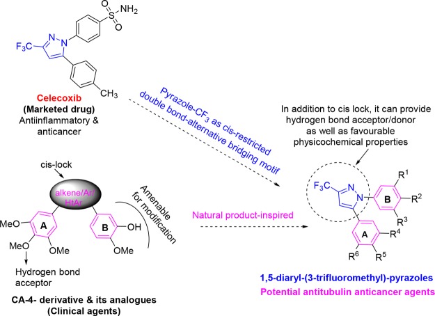
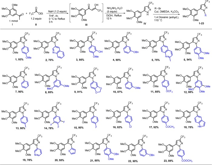
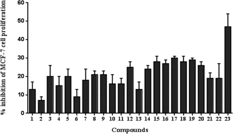
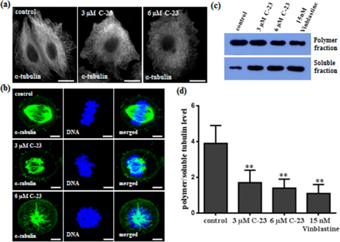
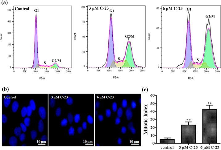
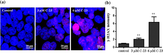
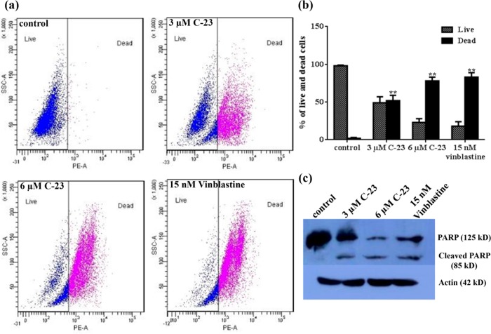
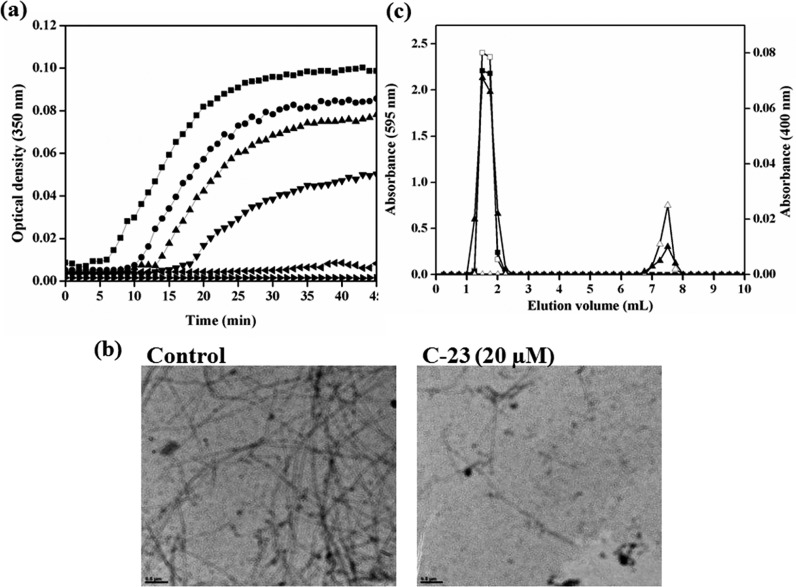
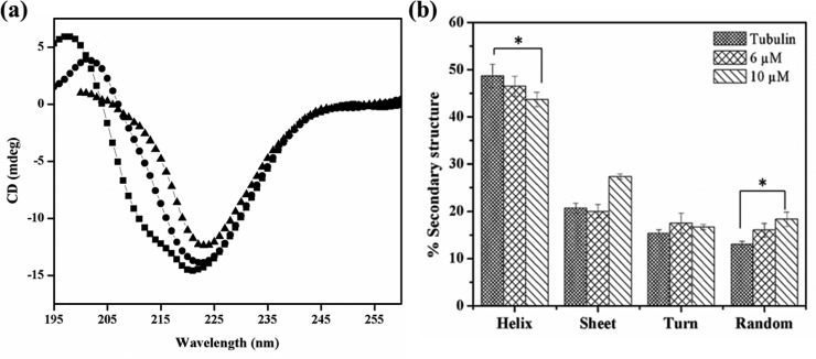
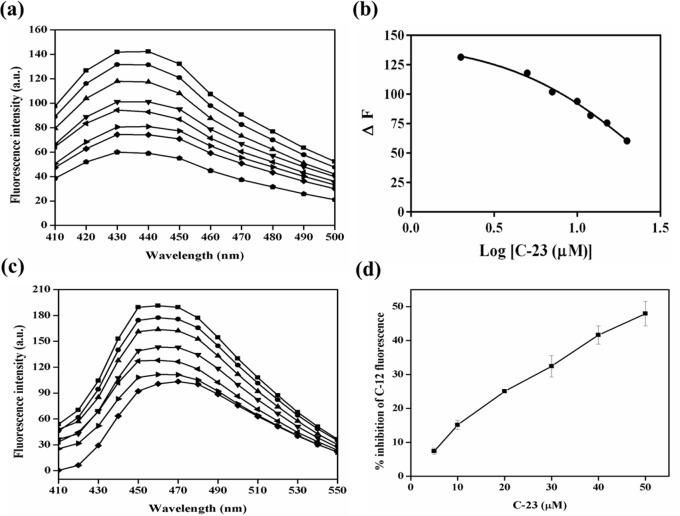
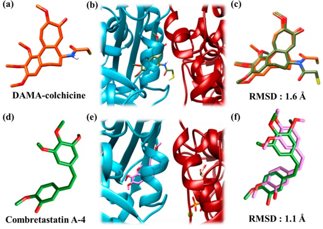
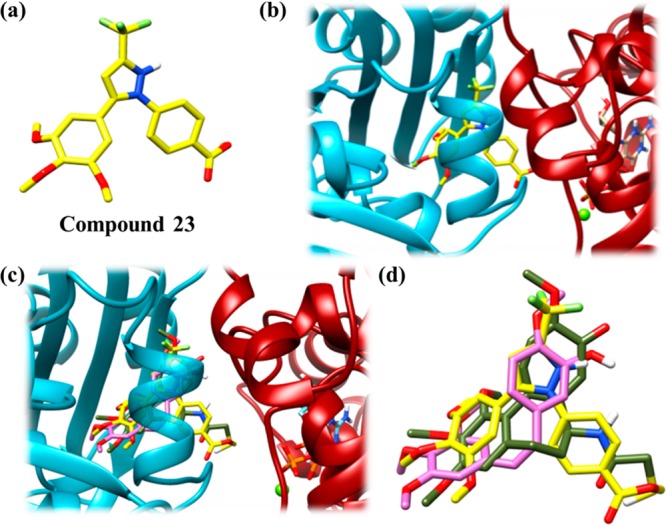
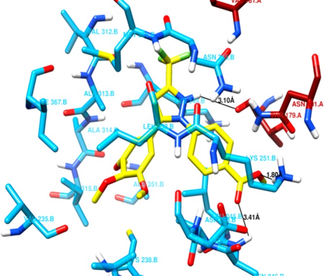
Twenty-three combretastatin A-4 (CA-4) analogues were synthesized by judiciously incorporating a functional N-heterocyclic motif present in Celecoxib (a marketed drug) while retaining essential pharmacophoric features of CA-4. Combretastatin-(trifluoromethyl)pyrazole hybrid analogues, i.e., 5-trimethoxyphenyl-3-(trifluoromethyl)pyrazoles with a variety of relevantly substituted aryls and heteroaryls at 1-position were considered as potential tubulin polymerization inhibitors. The cytotoxicity of the compounds was evaluated using MCF-7 cells. Analog 23 (C-23) was found to be the most active among the tested compounds. It showed pronounced cytotoxicity against HeLa, B16F10, and multidrug-resistant mammary tumor cells EMT6/AR1. Interestingly, C-23 displayed significantly lower toxicity toward noncancerous cells, MCF10A and L929, than their cancerous counterparts, MCF-7 and B16F10, respectively. C-23 depolymerized interphase microtubules, disrupted mitotic spindle formation, and arrested MCF-7 cells at mitosis, leading to cell death. C-23 inhibited the assembly of tubulin in vitro. C-23 bound to tubulin at the colchicine binding site and altered the secondary structures of tubulin. The data revealed the importance of (trimethoxyphenyl)(trifluoromethyl)pyrazole as a cis-restricted double bond-alternative bridging motif, and carboxymethyl-substituted phenyl as ring B for activities and interaction with tubulin. The results indicated that the combretastatin-(trifluoromethyl)pyrazole hybrid class of analogues has the potential for further development as anticancer agents.


Conflict of interest statement

The authors declare no competing financial interest.

Figures

Figure 1
Figure 1
Design of novel 1,5-diaryl-3-(trifluoromethyl)pyrazoles.
Figure 2
Figure 2
Synthesis of investigated compounds. Substrates, reagents, and conditions: compound IV (1 mmol), RBr (2equiv), CuI (5 mol %), DMEDA (20 mol %), K2CO3 (2.1 equiv), 1,4-dioxane (anhyd, 2 mL), 110 °C. Yield for maximum conversion in optimum time.
Figure 3
Figure 3
Screening of the antiproliferative activity of combretastatin analogues in MCF-7 cells. The percentage inhibition of MCF-7 cell proliferation by 1 μM CA-4 analogues was determined. The assay was performed four times. Error bar represents standard deviation.
Figure 4
Figure 4
C-23 depolymerized microtubules in MCF-7 cells. (a) C-23 depolymerized interphase microtubules in cells. Cells were incubated with vehicle (0.1% DMSO) and 3 and 6 μM C-23 for 36 h, fixed, and processed for immunostaining using α-tubulin IgG. The scale bar is 10 μm. (b) C-23 (3 and 6 μM) perturbed microtubule spindle formation in MCF-7 cells. DNA was stained with Hoechst 33258 (blue). The scale bar is 10 μm. (c) C-23 treatment leading to a decrease in the ratio of polymeric/soluble tubulin in MCF-7 cells. Cells were treated with vehicle (lane 1) and with 3 μM (lane 2) and 6 μM (lane 3) of C-23 for 36 h. Fifteen nanomolar vinblastine (lane 4) was used as a positive control. The experiment was performed four times. Shown is a representative blot. (d) Polymer and soluble level of tubulin quantified using ImageJ software and the ratio of polymeric/soluble tubulin was plotted. Error bar represents standard deviation. **p < 0.01 indicates statistical significance of the data.
Figure 5
Figure 5
C-23 blocked MCF-7 cells at mitosis. (a) Flow cytograms showing DNA distribution profiles of vehicle and C-23 (3 and 6 μM) treated MCF-7 cells in different phases of the cell cycle. (b) Effects of C-23 on mitotic progression. MCF-7 cells treated with vehicle and 3 and 6 μM C-23 for 36 h were fixed and DNA was stained with Hoechst 33258 (blue). The experiment was performed three times. (c) C-23 treatment increasing the mitotic index in MCF-7 cells. The experiment was performed three times and 500 cells were scored in each case. The error bar represents standard deviation. **p < 0.01 indicates statistical significance of the data.
Figure 6
Figure 6
C-23 induced DNA damage in cells. (a) MCF-7 cells were treated with vehicle and 3 and 6 μM C-23 for 36 h and were fixed and processed for immunostaining using γ-H2AX IgG to stain double-stranded DNA breaks (pink). DNA was stained using Hoechst 33258 (blue). The scale bar is 20 μm. (b) The γ-H2AX intensity was calculated using ImageJ software and plotted. The experiment was performed three times and 100 cells were scored for intensity calculation in each case. Error bar represents standard deviation. **p < 0.01 indicates statistical significance of the data.
Figure 7
Figure 7
C-23 treatment caused PARP cleavage and induced cell death in MCF-7 cells. (a) Flow cytograms show live and dead cells after PI staining. MCF-7 cells were incubated without and with C-23 (3 and 6 μM) for 48 h. Fifteen nanomolar vinblastine was used as a positive control. Representative images from three experiments are shown. (b) The percent of live and dead cells was quantified and plotted. The error bar shows standard deviation. **p < 0.01 indicates statistical significance of the data. (c) C-23 cleaves PARP in MCF-7 cells. Cells were treated with vehicle and 3 and 6 μM C-23 for 48 h. Cell lysate for each sample was prepared, and PARP cleavage was determined by Western blot using anti-PARP-1 IgG. Actin was used as a loading control. Fifteen nanomolar vinblastine was used as a positive control. The experiment was performed three times. A representative blot is shown.
Figure 8
Figure 8
C-23 bound to purified tubulin and inhibited its polymerization. (a) Tubulin (13 μM) was polymerized in the presence of vehicle (DMSO) (■) and 10 (●), 20 (▲), 40 (▼), 60 (◀), and 75 (▶) μM C-23. The kinetics of tubulin assembly was monitored at 350 nm. The experiment was performed three times. One of the independent sets is shown. (b) Electron micrographs of DMSO-induced tubulin polymers polymerized without (control) and with 20 μM C-23 are shown. The scale bar is 0.5 μm. (c)The elution profile of tubulin (20 μM) (□) and C-23 (60 μM) (Δ) when loaded individually onto the column are shown. Tubulin (20 μM) was incubated with C-23 (60 μM) in 25 mM PIPES at 25 °C for 30 min and then eluted through the same column. The elution profile of tubulin (■) and C-23 (▲) of the tubulin-C-23 complex is shown. The experiment was performed two times.
Figure 9
Figure 9
C-23 disrupted the secondary structure of purified tubulin. (a) Tubulin (1 μM) was incubated with vehicle (DMSO) (■) and 6 (●) and 10 (▲) μM C-23. The far-UV CD spectra were recorded. One of the three independent sets is shown. (b) The percentage of helix, sheet, turn, and random coil of tubulin when incubated without and with C-23 was determined by CDPro software and was plotted. The error bar indicates standard deviation. *p < 0.05 indicates statistical significance of the data.
Figure 10
Figure 10
C-23 bound at the colchicine binding site on tubulin. (a) Tubulin (5 μM) was incubated in the absence (■) and presence of 2 (●), 5 (▲), 7 (▼), 10 (◀), 12 (▶), 15 (◆), and 20 (⬟) μM C-23. Then the mixtures were incubated with 5 μM colchicine for 45 min at 37 °C. The fluorescence spectra (410–500 nm) were monitored using 350 nm as the excitation wavelength. (b) C-23 reduced the fluorescence intensity of tubulin–colchicine complex. One of the three independent sets is shown. (c) Tubulin (5 μM) was incubated in the absence (■) and presence of 5 (●), 10 (▲), 20 (▼), 30 (◀), 40 (▶), and 50 (◆) μM C-23. The mixtures were incubated with 5 μM C-12 for 10 min at 37 °C, and the fluorescence spectra (410–550 nm) were monitored using excitation wavelength 350 nm. One of the three independent sets is shown. (d) The percentage inhibition of the tubulin-C-12 fluorescence was plotted against C-23 concentration. The error bar indicates standard deviation.
Figure 11
Figure 11
DAMA-colchicine and CA-4 docked at the interface of the tubulin dimer. The color scheme for α-tubulin is dark red, for β-tubulin is cyan, for DAMA-colchicine (crystal structure) is orange red, for DAMA-colchicine (docked) is olive green, for CA-4 (crystal structure) is green, and for CA-4 (docked) is orchid pink. The compounds and tubulin are in stick and ribbon representations, respectively. Sulfur, hydrogen, oxygen, and nitrogen atoms are depicted as yellow, white, red, and dark blue sticks, respectively. (a) Crystal structure of DAMA-colchicine in sticks. (b) Docked conformation of DAMA-colchicine at the interface of the tubulin dimer. The conformation with lowest binding energy was found to be docked at the interface of the dimer. (c) RMS deviation between the crystal structure and docked conformation of DAMA-colchicine. The coordinates of the docked DAMA-colchicine (olive green) were superimposed over the X-ray crystallographically determined coordinates (orange red). (d) Crystal structure of CA-4 in sticks. (e) Docked conformation of CA-4 at the interface of tubulin dimer. The conformation of CA-4 with the lowest binding energy was found to be docked at the interface of the dimer. (f) RMS deviation between the docked and crystal structure of CA-4 obtained by superimposing the docked CA-4 (orchid pink) over the crystallographically determined coordinates (green).
Figure 12
Figure 12
C-23 docked at the colchicine binding pocket on the tubulin dimer. The color scheme for α-tubulin is dark red, for β-tubulin is cyan, for C-23 is yellow, for DAMA-colchicine (docked) is olive green and for CA-4 (docked) is orchid pink. The compounds and tubulin are in stick and ribbon representations, respectively. Hydrogen, oxygen, nitrogen, sulfur, and fluorine atoms are depicted as white, red, dark blue, yellow, and light green sticks, respectively. (a) Structure of C-23 in sticks. (b) Docked conformation of C-23 at the interface of tubulin dimer. The conformation of C-23 with the lowest binding energy was found to be docked at the interface of the dimer (c) Docked conformation of C-23 overlapped with docked conformations of DAMA-colchicine and CA-4. All the three compounds were shown to occupy the same binding pocket. (d) Zoomed view of (c) showing overlapped C-23, CA-4, and DAMA-colchicine.
Figure 13
Figure 13
Amino acid residues of the tubulin dimer present within 4 Å distance of C-23. Residues of α-tubulin and β-tubulin are shown in dark red and cyan sticks, respectively. C-23 is in yellow stick representation. The color scheme for the atoms is same as in Figure 12. The black line represents the possibility of a hydrogen bond between C-23 and the amino acid present in its binding pocket. C-23 was found to have possible hydrogen bonds with Thr179A (3.10 Å), Asp248B (3.41 Å), and Lys251B (1.80 Å).

References

    1. Jordan M. A.; Wilson L. Microtubules as a Target for Anticancer Drugs. Nat. Rev. Cancer 2004, 4 (4), 253–265. 10.1038/nrc1317. - DOI - PubMed
    1. Field J. J.; Kanakkanthara A.; Miller J. H. Microtubule-Targeting Agents Are Clinically Successful due to Both Mitotic and Interphase Impairment of Microtubule Function. Bioorg. Med. Chem. 2014, 22 (18), 5050–5059. 10.1016/j.bmc.2014.02.035. - DOI - PubMed
    1. Singh P.; Rathinasamy K.; Mohan R.; Panda D. Microtubule Assembly Dynamics: An Attractive Target for Anticancer Drugs. IUBMB Life 2008, 60 (6), 368–375. 10.1002/iub.42. - DOI - PubMed
    1. Dumontet C.; Jordan M. A. Microtubule-Binding Agents: A Dynamic Field of Cancer Therapeutics. Nat. Rev. Drug Discovery 2010, 9 (10), 790–803. 10.1038/nrd3253. - DOI - PMC - PubMed
    1. Kanthou C.; Tozer G. M. Microtubule Depolymerizing Vascular Disrupting Agents: Novel Therapeutic Agents for Oncology and Other Pathologies. Int. J. Exp. Pathol. 2009, 90 (3), 284–294. 10.1111/j.1365-2613.2009.00651.x. - DOI - PMC - PubMed