. 2018 Apr 30;3(4):4466-4474.
doi: 10.1021/acsomega.8b00566. Epub 2018 Apr 24.

Fully Conjugated Porphyrin Glass: Collective Light-Harvesting Antenna for Near-Infrared Fluorescence beyond 1 μm

Affiliations

Fully Conjugated Porphyrin Glass: Collective Light-Harvesting Antenna for Near-Infrared Fluorescence beyond 1 μm

Mitsuhiko Morisue et al. ACS Omega. .

Abstract

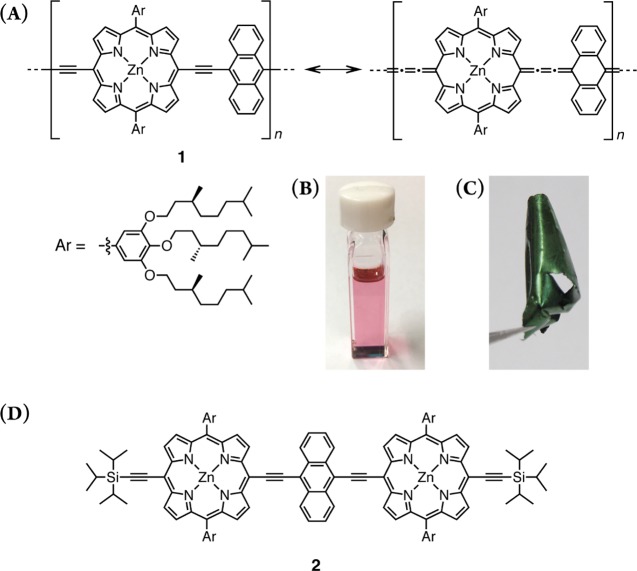
Expanded π-systems with a narrow highest occupied molecular orbital-lowest unoccupied molecular orbital band gap encounter deactivation of excitons due to the "energy gap law" and undesired aggregation. This dilemma generally thwarts the near-infrared (NIR) luminescence of organic π-systems. A sophisticated cofacially stacked π-system is known to involve exponentially tailed disorder, which displays exceptionally red-shifted fluorescence even as only a marginal emission component. Enhancement of the tail-state fluorescence might be advantageous to achieve NIR photoluminescence with an expected collective light-harvesting antenna effect as follows: (i) efficient light-harvesting capacity due to intense electronic absorption, (ii) a long-distance exciton migration into the tail state based on a high spatial density of the chromophore site, and (iii) substantial transmission of NIR emission to circumvent the inner filter effect. Suppression of aggregation-induced quenching of fluorescence could realize collective light-harvesting antenna for NIR-luminescence materials. This study discloses an enhanced tail-state NIR fluorescence of a self-standing porphyrin film at 1138 nm with a moderate quantum efficiency based on a fully π-conjugated porphyrin that adopts an amorphous form, called "porphyrin glass".


Conflict of interest statement

The authors declare no competing financial interest.

Figures

Figure 1
Figure 1
Schematic representation of the density of states and the corresponding fluorescence spectrum as the sum of Gaussian statistics (black solid lines) and exponential tails (red dotted line) (A) and the space coordinate of the occupied density of states (B).
Scheme 1
Scheme 1. Synthesis of 2
Figure 2
Figure 2
Chemical structure of 1 in a benzenoid–acetylene form and a quinoid–cumulene form (A), and photographs of the toluene solution in a cuvette (B) and the porphyrin foil (C) under ambient light. Chemical structure of a model compound 2 (D).
Figure 3
Figure 3
(A) Electronic absorption spectra of 1 at 298 K in toluene (red line) and spectrometric titration with pyridine (gray and green lines). (B) Fluorescence spectra of 1 in toluene under argon-saturated conditions obtained by excitation at 450 nm in the absence and presence of pyridine (red and green, respectively), together with normalized excitation spectra monitored at 800 nm (gray line) and 1000 nm (black line) in the absence of pyridine.
Figure 4
Figure 4
Spectrometric titration of 2 ([2]0 = 1.4 × 10⁻⁴ M, red line) with pyridine (gray lines up to 1 × 10² equivalent, and 1 × 10⁴ equivalent, green line) in electronic absorption and fluorescence spectra at 25 °C in toluene (A). Stepwise binding constants were estimated as K1 = 4.1 × 10⁴ M–1 and K2 = 8.3 × 10³ M–1 based on global titration analyses (B).
Figure 5
Figure 5
Possible Jablonski diagram, combined with the electronic absorption spectrum deconvoluted with Gaussian-curve fitting (yellow) and the fluorescence spectrum (green) in toluene.
Figure 6
Figure 6
Electronic absorption (black), fluorescence (red), and excitation (gray) spectra of a spin-cast film of 2 on a quartz substrate. Emission and excitation spectra were obtained by excitation at 443 nm and monitoring at 958 nm, respectively.
Figure 7
Figure 7
Emission–excitation contour map of the porphyrin foil. Excitation spectrum monitored at 1000 nm (black line), absorption spectrum obtained by spectroscopic ellipsometry (gray line, adapted from ref (28)) (left panel), and emission spectra obtained by excitation at 470 and 800 nm (upper panel). It should be noticed that no emission at 827 nm was found.
Figure 8
Figure 8
Possible Jablonski diagram, combined with the electronic absorption spectrum in toluene (yellow) and the fluorescence spectrum of porphyrin foil (red).
Figure 9
Figure 9
WAXD profile of the porphyrin foil (red line, adapted from ref (28)) and GIWAXD profile of the spin-cast film of the porphyrin array on a silicon wafer (blue line) after background correction (A), where representative peaks are shown as d-spacing (d = 2π/q). The observed GIWAXD pattern (B) and azimuthal-angle dependence of the intensity of the primary GIWAXD peak at the magnitude of the scattering vector, q, of approximately 2.1 nm–1 (C).
Figure 10
Figure 10
Geometry-optimized models of the bundle structure of π-stacked chains (blue and red chains) (A), and distorted single backbone (B) produced using an MM+ force field (HyperChem Ver. 8.0 software).

References

    1. Smith A. M.; Mancini M. C.; Nie S. Second window for in vivo imaging. Nat. Nanotechnol. 2009, 4, 710–711. 10.1038/nnano.2009.326. - DOI - PMC - PubMed
    1. Hong G.; Antaris A. L.; Dai H. Near-infrared fluorophores for biomedical imaging. Nat. Biomed. Eng. 2017, 1, 001010.1038/s41551-016-0010. - DOI
    1. Schnermann M. J. Organic dyes for deep bioimaging. Nature 2017, 551, 176–177. 10.1038/nature24755. - DOI - PubMed
    1. Liang Y.; Yu L. Bulk heterojunction solar cells with exceptionally high performance. Acc. Chem. Res. 2010, 43, 1227–1236. 10.1021/ar1000296. - DOI - PubMed
    1. Dou L.; Liu Y.; Hong Z.; Li G.; Yang Y. Low-bandgap near-IR conjugated polymer/molecules for organic electronics. Chem. Rev. 2015, 115, 12633–12655. 10.1021/acs.chemrev.5b00165. - DOI - PubMed