. 2018 Jun 30;3(6):5980-5987.
doi: 10.1021/acsomega.8b00347. Epub 2018 Jun 4.

Self-Assembly of a Dentinogenic Peptide Hydrogel

Affiliations

Self-Assembly of a Dentinogenic Peptide Hydrogel

Peter K Nguyen et al. ACS Omega. .

Abstract

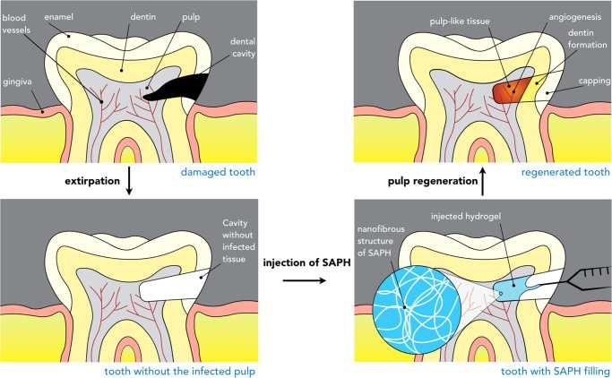
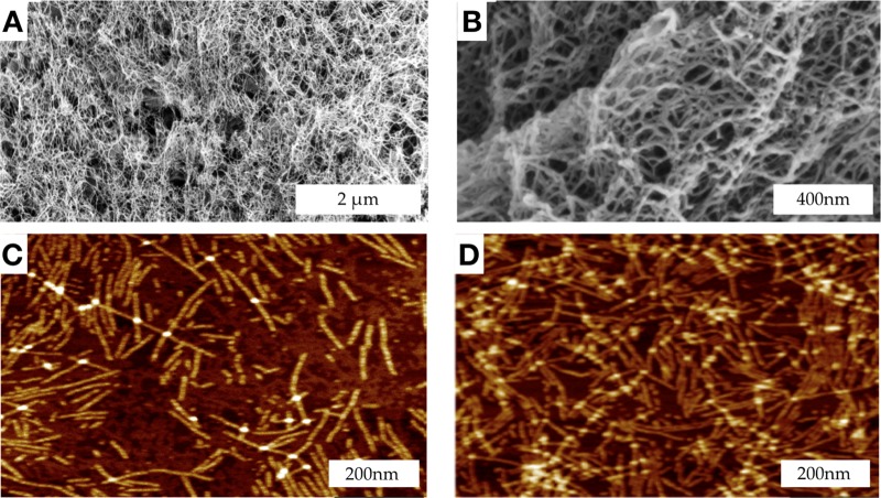
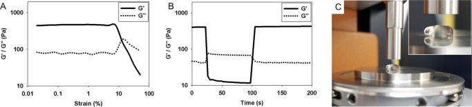
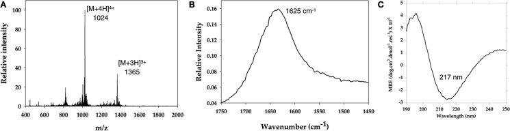
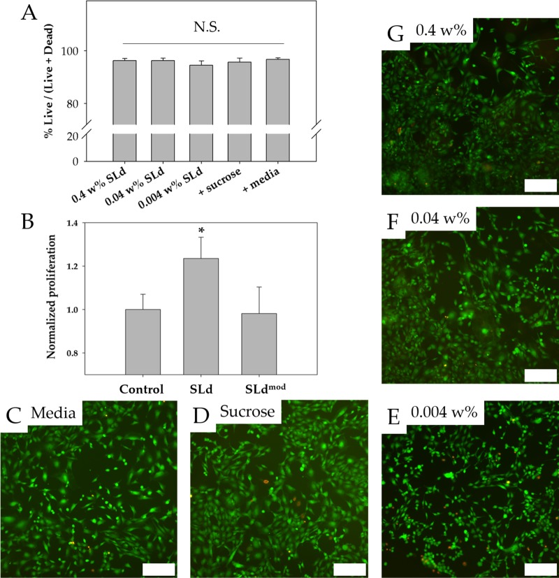
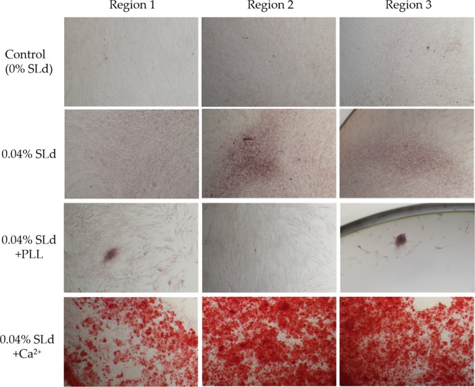
Current standard of care for treating infected dental pulp, root canal therapy, retains the physical properties of the tooth to a large extent, but does not aim to rejuvenate the pulp tissue. Tissue-engineered acellular biomimetic hydrogels have great potential to facilitate the regeneration of the tissue through the recruitment of autologous stem cells. We propose the use of a dentinogenic peptide that self-assembles into β-sheet-based nanofibers that constitute a biodegradable and injectable hydrogel for support of dental pulp stem cells. The peptide backbone contains a β-sheet-forming segment and a matrix extracellular phosphoglycoprotein mimic sequence at the C-terminus. The high epitope presentation of the functional moiety in the self-assembled nanofibers may enable recapitulation of a functional niche for the survival and proliferation of autologous cells. We elucidated the hierarchical self-assembly of the peptide through biophysical techniques, including scanning electron microscopy and atomic force microscopy. The material property of the self-assembled hydrogel was probed though oscillatory rheometry, demonstrating its thixotropic nature. We also demonstrate the cytocompatibility of the hydrogel with respect to fibroblasts and dental pulp stem cells. The self-assembled peptide platform holds promise for guided dentinogenesis and it can be tailored to a variety of applications in soft tissue engineering and translational medicine in the future.


Conflict of interest statement

The authors declare no competing financial interest.

Figures

Scheme 1
Scheme 1. Peptide Self-Assembly and Gross Hydrogel Structure
(A–C) The base sequence has alternative hydrophilic and hydrophobic residues. In aqueous media, the hydrophobic leucine residues collapse into a core, exposing the hydrophilic serine residues to the surface. Stacking of a tetrameric unit to maximize hydrogen bonding leads to the formation of a β-sheet nanofiber. The lengthening and crosslinking of the nanofibers is favored in physiological salt concentrations as the repulsion among the positively charged lysine residues is shielded, leading to (D, E) the formation of robust hydrogels that maintain their shape.
Scheme 2
Scheme 2. Injectable Peptide Scaffolds for Pulpal Regeneration
The dental cavity may contain bacterial colonies that need to be extirpated (partial or complete pulpotomy). After clinical extirpation, the void may be filled with the thixotropic peptide hydrogel with dentinogenic activity. As the hydrogel promptly recovers after shear thinning, it should reassemble into a viscoelastic scaffold, filling the void. Recruitment and differentiation of autologous stem cells may then lead to re-establishment of a pulplike tissue niche.
Figure 1
Figure 1
Ultrastructure of the hydrogel formed by SLd. Critical point-dried hydrogel samples in SEM show formation of a dense nanofibrous network with nanoscale pores (A) at low magnification (50k×) and (B) at high magnification (200k×). (C, D) Drop-cast samples of diluted SLd on two-dimensional mica disks show characteristic nanofibrous structure with individual fibers visible in peak force AFM. Measurement of fiber dimensions showed a width of ∼14 nm and a height of ∼2 nm, which is consistent with the proposed self-assembly scenario in Scheme 1.
Figure 2
Figure 2
Rheological characterization of hydrogels to demonstrate thixotropy and injectability. (A) Strain sweep of peptide hydrogels show a relatively high storage modulus (G′) of about 400 Pa, which is decimated above ∼10% strain. At this point the values for G″ exceed G′ indicating liquefaction. (B) Hydrogels are allowed to equilibrate at a constant frequency of 1 Hz and 1% strain rate. As the strain is instantaneously increased to 100%, inversions of G′ and G″ occur suggesting instantaneous liquefaction of the gel. Interestingly, after removing the high deformation strain, 100% of the storage modulus is recovered within 3 s. (C) This is evident while pipetting the gel onto the rheometer stage where instantaneous gelation leaves a nubbin suspended with its reflection visible.
Figure 3
Figure 3
Spectroscopic characterization of the dentinogenic hybrid peptide. (A) Characterization of SLd via mass spectroscopy shows expected [M + 3H]3+ and [M + 4H]4+ peaks at 1365 and 1024 Da, consistent with the molecular weight (4095 Da). (B) The FTIR spectrum demonstrates the characteristic β-sheet signature peak at 1625 cm–1. (C) Circular dichroism spectrum contains a minimum at 217 nm, which is specific to a β-sheet secondary structure.
Figure 4
Figure 4
Cytocompatibility and DPSC proliferation. (A) Viability of fibroblasts cultured with SLd at different concentrations (0.004–0.4 wt % w/v), media with formulation solution (298 mM sucrose), or media-only are shown along with their representative live/dead images (C–G). (B) Dental pulp stem cells demonstrated stronger proliferation (CCK-8) compared to no treatment of 0.004 wt % SLd or treatment with a modified variant (SLdmod) with similar charge density (n = 8, *p < 0.01). Scale bar (C–G): 200 μm.
Figure 5
Figure 5
Calcium phosphate deposition in response to SLd treatment of DPSCs (representative regions from independent cultures). SLd hydrogel shows appreciable red staining either alone or in combination with poly-l-lysine (PLL) compared to media-only controls. The addition of Ca2+ to the SLd formulation results in significantly more observable calcium phosphate deposition, as observed with Alizarin red staining.

References

    1. Dye B.; Thornton-Evans G.; Li X.; Iafolla T.. Dental Caries and Tooth Loss in Adults in the United States, 2011–2012. In NCHS Data Brief No. 197; National Center for Health Statistics: Hyattsville, MD, 2015. - PubMed
    1. Gulabivala K.; Ng Y. L.. 1 - Tooth Organogenesis, Morphology and Physiology. In Endodontics, 4th ed.; Mosby, 2014; pp 2–32.
    1. Ostby B. N. The role of the blood clot in endodontic therapy. An experimental histologic study. Acta Odontol. Scand. 1961, 19, 324–353. 10.3109/00016356109043395. - DOI - PubMed
    1. Myers W. C.; Fountain S. B. Dental pulp regeneration aided by blood and blood substitutes after experimentally induced periapical infection. Oral Surg., Oral Med., Oral Pathol. 1974, 37, 441–50. 10.1016/0030-4220(74)90119-4. - DOI - PubMed
    1. Huang G. T. Pulp and dentin tissue engineering and regeneration: current progress. Regener. Med. 2009, 4, 697–707. 10.2217/rme.09.45. - DOI - PMC - PubMed

LinkOut - more resources