. 2018 Oct 1;128(10):4485-4500.
doi: 10.1172/JCI99768. Epub 2018 Jul 19.

ASK1 contributes to fibrosis and dysfunction in models of kidney disease

Affiliations

ASK1 contributes to fibrosis and dysfunction in models of kidney disease

John T Liles et al. J Clin Invest. .

Abstract

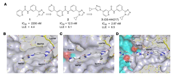
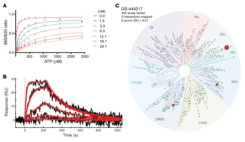
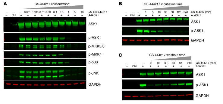
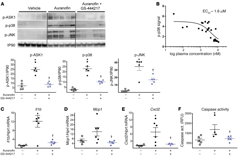
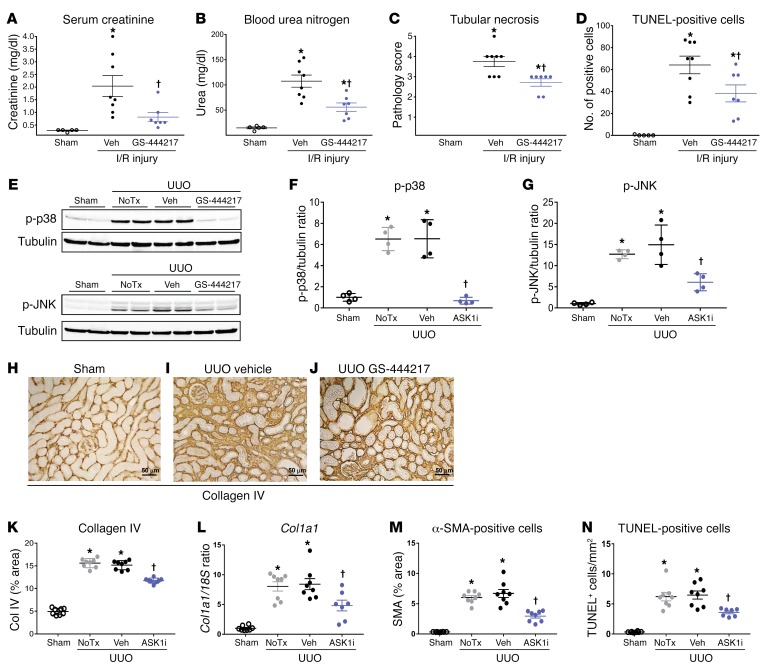
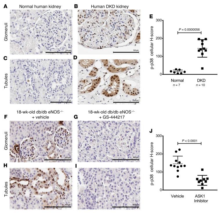
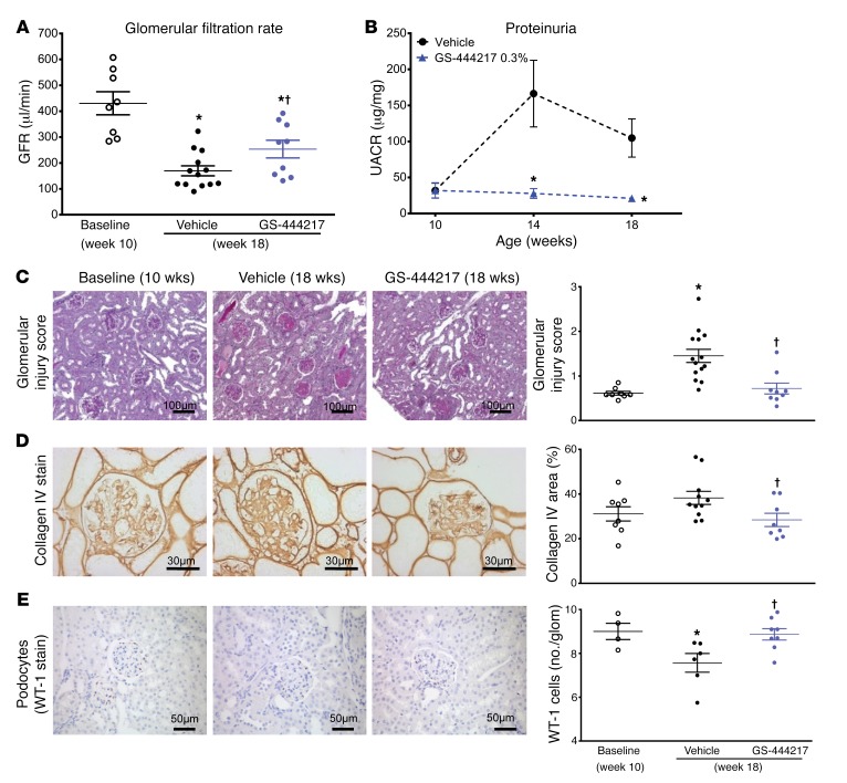
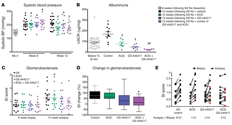
Oxidative stress is an underlying component of acute and chronic kidney disease. Apoptosis signal-regulating kinase 1 (ASK1) is a widely expressed redox-sensitive serine threonine kinase that activates p38 and c-Jun N-terminal kinase (JNK) mitogen-activated protein kinase kinases, and induces apoptotic, inflammatory, and fibrotic signaling in settings of oxidative stress. We describe the discovery and characterization of a potent and selective small-molecule inhibitor of ASK1, GS-444217, and demonstrate the therapeutic potential of ASK1 inhibition to reduce kidney injury and fibrosis. Activation of the ASK1 pathway in glomerular and tubular compartments was confirmed in renal biopsies from patients with diabetic kidney disease (DKD) and was decreased by GS-444217 in several rodent models of kidney injury and fibrosis that collectively represented the hallmarks of DKD pathology. Treatment with GS-444217 reduced progressive inflammation and fibrosis in the kidney and halted glomerular filtration rate decline. Combination of GS-444217 with enalapril, an angiotensin-converting enzyme inhibitor, led to a greater reduction in proteinuria and regression of glomerulosclerosis. These results identify ASK1 as an important target for renal disease and support the clinical development of an ASK1 inhibitor for the treatment of DKD.

Keywords: Apoptosis; Chronic kidney disease; Fibrosis; Nephrology; Therapeutics.


Conflict of interest statement

Conflict of interest: JTL, BKC, GTN, EBL, GB, FHK, SSB, ML, SP, BS, SW, DF, TS, EH, MW, GAP, MG, and DGB are or were employees and/or shareholders of Gilead Sciences. LC, KAK, FYM, DJNP, TA, HY, and ABF received research reagents or financial support to conduct studies for Gilead Sciences.

Figures

Figure 1
Figure 1. Structure-based drug design of compound 3, GS-444217.
(A) Medicinal chemistry optimization of compound 1 showing improved biochemical potency (IC50) and maintenance of low lipophilicity resulting in improved lipophilic ligand efficiency (LLE). (B) Structure of compound 1 occupying the ATP-binding pocket of ASK1. Interactions between hinge residue Val757 and the catalytic residue Lys709 are highlighted. (C) Structure of compound 2 bound to ASK1 with a cross-dimer interaction to the Tyr814 residue of the neighboring ASK1 monomer. (D) Structure of compound 3 (GS-444217) bound to ASK1.
Figure 2
Figure 2. GS-444217, a selective ATP-competitive inhibitor of ASK1.
(A) Enzymatic competition of GS-444217 with ATP was measured in the presence of increasing concentrations of GS-444217 (0–24 nM) and ATP (0–2,400 μM). (B) Kinetics and binding affinity of GS-444217 to ASK1 using surface plasmon resonance. Black traces represent the experimental data, and red traces represent the fit of a simple 1:1 interaction model to the data. (C) Selectivity of GS-444217 (1 μM) for ASK1 was determined using KINOMEscan binding assay against 442 kinases. Map describes quantitatively the interaction patterns of GS-444217 with major kinase families: tyrosine kinases (TK), tyrosine kinase–like (TKL), serine/threonine kinases (STE), casein kinase 1 (CK1), protein kinases A, G, C (AGC), calmodulin/calcium-regulated kinases (CAMK), CDK/MAPK/GSK3/CLK (CMGC), and Other, consisting of many diverse families. The sphere radius corresponds to inhibitor affinity. Size of red dots shows relative affinity of GS-444217 for ASK1 (largest dot), ribosomal s6 kinase-4 (RSK4), and dual-specificity tyrosine phosphorylation–regulated kinase-1A (DYRK1A).
Figure 3
Figure 3. GS-444217 dose-dependently inhibits ASK1 activity.
ASK1 inhibition assays using HEK293T cells with adenoviral overexpression of human ASK1 (AdASK1). (A) Cells were treated for 2 hours with 1:3 dilutions of GS-444217 (0.001–10 μM). Phosphorylation of ASK1 and ASK1 substrates (MKK3/6, MKK4, and their respective downstream substrates p38 and JNK kinases) was measured by Western blot. GAPDH was used as a protein-loading control. (B) Time course of ASK1 inhibition following treatment of cells with 1 μM GS-444217 (treatment duration of 1 minute to 4 hours). (C) Reversibility of ASK1 inhibition following washout of GS-444217 and replacement with serum-free media (washout time of 0–240 minutes).
Figure 4
Figure 4. GS-444217 inhibits activation of ASK1, p38, and JNK in rat kidney.
(A and CF) A single oral dose of GS-444217 (30 mg/kg, n = 8) or vehicle (equal volume, n = 5) was administered to Sprague-Dawley rats, which were challenged 30 minutes later with auranofin (30 mg/kg, i.p.) to cause OS-induced activation of the ASK1 pathway. Kidney cortex samples were collected 30 minutes after auranofin administration. (A) Western blot analysis of renal cortex lysates showing p-ASK1, p-p38, and p-JNK levels after in vivo administration of vehicle, auranofin, or auranofin and 30 mg/kg GS-444217. Dot plots show p-ASK1, p-p38, and p-JNK levels normalized to IP90 loading control. (B) GS-444217 (10 mg/kg, n = 14, or 30 mg/kg, n = 12, p.o.) was administered to Sprague-Dawley rats. Plasma was collected from individual rats over the course of the dosing interval, and kidneys were collected at the end of the time course. Based on plasma concentrations of GS-444217 and the corresponding p-p38 signal in each kidney (measured by ELISA in renal cortex lysates), the in vivo EC50 of GS-444217 for inhibition of the renal ASK1 pathway was estimated to be 1.6 μM. (CF) Relative mRNA expression of inflammatory cytokines (Il1b, Ccl2, and Cxcl2) and caspase activity were measured in kidneys of rats treated with vehicle, auranofin, or auranofin and 30 mg/kg GS-444217. RFU, relative fluorescence units. For A and CF, data are mean ± SEM; *P < 0.01 vs. control, P < 0.01 vs. auranofin (ANOVA with Bonferroni’s multiple-comparisons test).
Figure 5
Figure 5. GS-444217 inhibits acute renal tubular injury in rat kidney.
(AD) Renal ischemia/reperfusion (I/R) injury. GS-444217 (30 mg/kg) or vehicle (Veh; equal volume) was orally administered to Sprague-Dawley rats just before 30 minutes of bilateral renal ischemia. Parameters of renal function were assessed in serum and kidney following a 24-hour reperfusion period. (A and B) Serum creatinine and blood urea nitrogen concentrations. (C and D) Renal pathology scores for tubular necrosis (H&E-stained sections) and apoptosis/necrosis (TUNEL stain). Data in AD are mean ± SEM, n = 5–8; *P < 0.05 vs. control, P < 0.05 vs. I/R treated with vehicle (ANOVA with Bonferroni’s multiple-comparisons test). (EN) Unilateral ureteral obstruction (UUO). Sprague-Dawley rats had sham or UUO surgery. GS-444217 (30 mg/kg) or vehicle (equal volume) was orally administered 1 hour before surgery and continued twice per day for 7 days. (EG) Western blot analysis of kidney lysates for p-p38 and p-JNK. Dot plots show p-p38 and p-JNK levels normalized to tubulin loading control (NoTx, no treatment). (HK, M, and N) Image analysis and graphed pathology scores of renal sections stained for collagen deposition (collagen IV) (scale bars: 50 μm) (HK), cortical interstitial α-smooth muscle actin–positive (α-SMA–positive) myofibroblasts (M), and apoptosis/necrosis of kidney epithelial cells (TUNEL) (N). (L) Collagen I (Col1a1) mRNA was measured in whole kidney by reverse transcriptase PCR. Data in F, G, and KN are mean ± SEM, n = 4 (sham), n = 8 (UUO); *P < 0.01 vs. sham surgery, P < 0.01 vs. UUO treated with vehicle (ANOVA with Bonferroni’s multiple-comparisons test).
Figure 6
Figure 6. The ASK1 pathway is activated similarly in DKD patients and db/db eNOS–/– mice.
(AJ) Immunostaining for p-p38 was used as a measure of ASK1 pathway activation in kidney biopsies from donors without kidney disease (“normal,” n = 7) (A and C) and patients with DKD (n = 10) (B and D), and kidneys from 18-week-old db/db eNOS–/– mice treated with vehicle (standard rodent chow) (F and H) or with GS-444217 (0.3% in rodent chow) (G and I) for 8 weeks (n = 8–10). Representative images of glomerular (top panels) and tubulointerstitial compartments (bottom panels) are shown for human and mouse (scale bars: 100 μm). (E and J) Whole-slide images were analyzed with Definiens Developer XD and expressed as an H-Score that quantifies p-p38 staining intensity and distribution in kidney biopsies from DKD patients and controls (E) and in kidneys from 18-week-old db/db eNOS–/– mice treated with vehicle or with GS-444217 for 8 weeks (J) (mean ± SD; P value is shown on graph; unpaired t test).
Figure 7
Figure 7. GS-444217 halts the progressive decline of renal function in a mouse model of DKD.
(AE) Ten-week-old db/db eNOS–/– mice were fed standard rodent chow (vehicle) or chow containing GS-444217 (0.3% by weight) for 8 weeks. A separate group of untreated db/db eNOS–/– mice were euthanized at 10 weeks of age to establish baseline-disease parameters. (A) GFR was measured using inulin-FITC clearance in baseline mice (week 10) and in treated mice (week 18). (B) Proteinuria (urinary albumin to creatinine ratio [UACR]) was measured at weeks 10, 14, and 18. (C) A sclerosis injury score was calculated at 18 weeks on periodic acid–Schiff–stained kidneys averaging the lesion scores of 80–100 glomeruli per mouse using the following scoring system: 0, no lesion; 1, sclerosis of up to 25% of the glomerulus; 2, sclerosis of 25%–50%; 3, sclerosis of 50%–75%; and 4, sclerosis of >75% of the glomerulus. (D and E) Image analysis and graphed pathology scores of renal sections stained for collagen deposition (collagen IV) (D) or podocyte loss (Wilms tumor antigen [WT-1]) (E). Scale bars: 100 μm (C), 50 μm (E), or 30 μm (D). Data in AE are mean ± SEM, n = 9–12; *P < 0.05 vs. 10-week baseline by unpaired t test, P < 0.05 vs. vehicle (ANOVA with Bonferroni’s multiple-comparisons test).
Figure 8
Figure 8. GS-444217 increases the efficacy of enalapril in a chronic glomerular injury model.
(AE) Eight weeks after 5/6 nephrectomy (5/6 Nx), Sprague-Dawley rats were randomized into groups based on sclerosis index (SI) scores determined from kidney biopsies and fed standard rodent chow (control), chow containing GS-444217 (0.3% by weight plus 30 mg/kg, once per day, p.o.), the angiotensin-converting enzyme inhibitor (ACEi) enalapril (50 mg/l in drinking water), or enalapril with GS-444217 for 4 weeks. (A) Systolic blood pressure (BP) was measured at week 0 (gray dots) and 8 and 12 weeks after Nx (black dots, control; green dots, ACEi; blue dots, GS-444217; purple dots, ACEi plus GS-444217). (B) Urinary albumin to creatinine ratio (UACR) before treatment (gray dots, averaged for all groups 8 weeks after Nx) or 4 weeks after Nx and treatment. (CE) Glomerulosclerosis severity was measured by biopsy (8 weeks after Nx) or on whole kidney sections (12 weeks after Nx). (C) SI score was calculated on periodic acid–Schiff–stained kidney sections averaging the lesion scores of 80–100 glomeruli per rat (scoring system: 0, no lesion; 1, sclerosis of up to 25% of the glomerulus; 2, sclerosis of 25%–50%; 3, sclerosis of 50%–75%; 4, sclerosis of >75%). (D) Progression of glomerulosclerosis from week 8 to week 12 (box and whisker plot, minimum to maximum). (E) Before-and-after plots of SI scores at 8 weeks after Nx (biopsy; circles) and 12 weeks after Nx (autopsy; squares). Red squares indicate which rats showed regression of glomerulosclerosis from biopsy to autopsy. Numbers of rats (red font) per group in which glomerulosclerosis regressed are shown below the x axis. Data in AE are mean ± SEM, n = 12–14; *P < 0.05 vs. 8-week baseline by unpaired t test, P < 0.05 vs. vehicle (ANOVA with Bonferroni’s multiple-comparisons test); #P < 0.01 vs. GS-444217 or ACEi monotherapy (unpaired t test).

Comment in

References

    1. Breyer MD, Susztak K. The next generation of therapeutics for chronic kidney disease. Nat Rev Drug Discov. 2016;15(8):568–588. doi: 10.1038/nrd.2016.67. - DOI - PMC - PubMed
    1. Fernandez-Fernandez B, Ortiz A, Gomez-Guerrero C, Egido J. Therapeutic approaches to diabetic nephropathy — beyond the RAS. Nat Rev Nephrol. 2014;10(6):325–346. doi: 10.1038/nrneph.2014.74. - DOI - PubMed
    1. Evans M, Bain SC, Hogan S, Bilous RW, Collaborative Study Group participants Irbesartan delays progression of nephropathy as measured by estimated glomerular filtration rate: post hoc analysis of the Irbesartan Diabetic Nephropathy Trial. Nephrol Dial Transplant. 2012;27(6):2255–2263. doi: 10.1093/ndt/gfr696. - DOI - PubMed
    1. Reidy K, Kang HM, Hostetter T, Susztak K. Molecular mechanisms of diabetic kidney disease. J Clin Invest. 2014;124(6):2333–2340. doi: 10.1172/JCI72271. - DOI - PMC - PubMed
    1. Najafian B, Alpers CE, Fogo AB. Pathology of human diabetic nephropathy. Contrib Nephrol. 2011;170:36–47. doi: 10.1159/000324942. - DOI - PubMed

Publication types

MeSH terms

Substances