. 2018 Oct 1;128(10):4397-4412.
doi: 10.1172/JCI99436. Epub 2018 Jul 19.

Specific covalent inhibition of MALT1 paracaspase suppresses B cell lymphoma growth

Affiliations

Specific covalent inhibition of MALT1 paracaspase suppresses B cell lymphoma growth

Lorena Fontán et al. J Clin Invest. .

Abstract

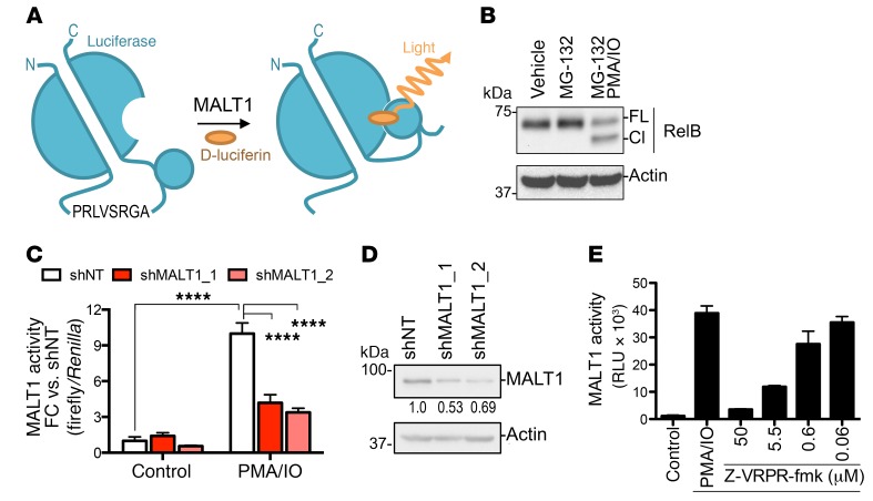
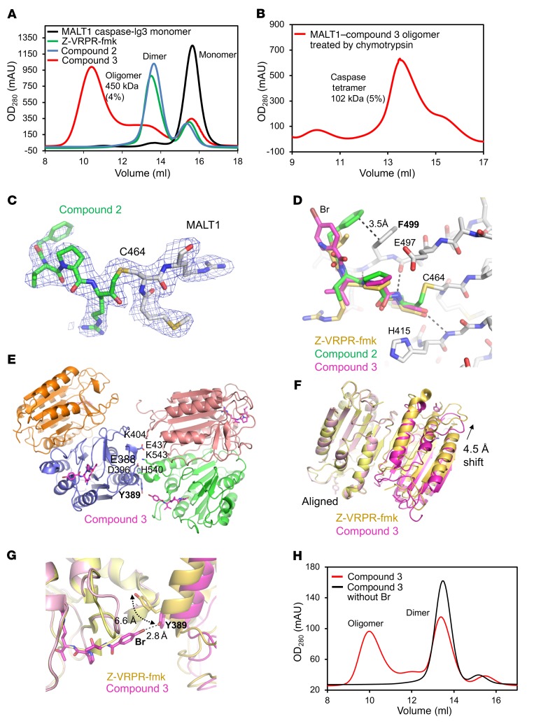
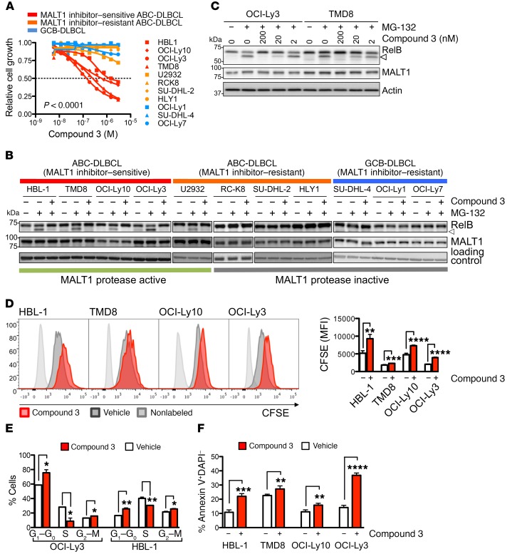
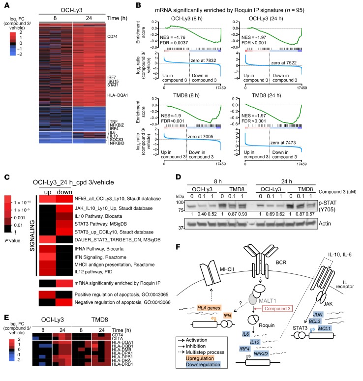
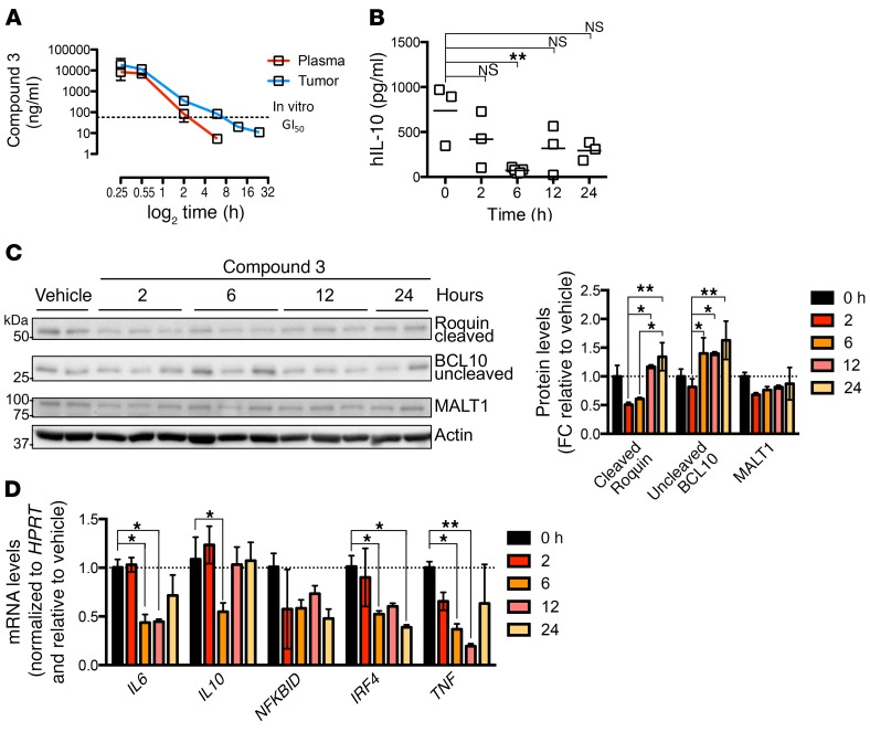
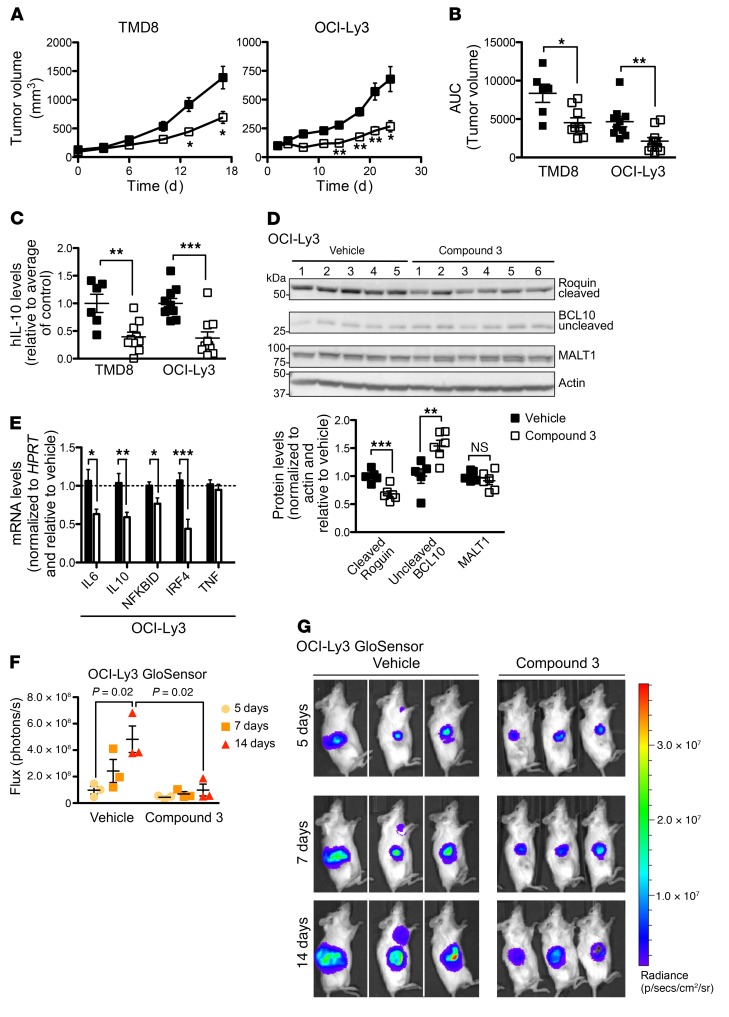
The paracaspase MALT1 plays an essential role in activated B cell-like diffuse large B cell lymphoma (ABC DLBCL) downstream of B cell and TLR pathway genes mutated in these tumors. Although MALT1 is considered a compelling therapeutic target, the development of tractable and specific MALT1 protease inhibitors has thus far been elusive. Here, we developed a target engagement assay that provides a quantitative readout for specific MALT1-inhibitory effects in living cells. This enabled a structure-guided medicinal chemistry effort culminating in the discovery of pharmacologically tractable, irreversible substrate-mimetic compounds that bind the MALT1 active site. We confirmed that MALT1 targeting with compound 3 is effective at suppressing ABC DLBCL cells in vitro and in vivo. We show that a reduction in serum IL-10 levels exquisitely correlates with the drug pharmacokinetics and degree of MALT1 inhibition in vitro and in vivo and could constitute a useful pharmacodynamic biomarker to evaluate these compounds in clinical trials. Compound 3 revealed insights into the biology of MALT1 in ABC DLBCL, such as the role of MALT1 in driving JAK/STAT signaling and suppressing the type I IFN response and MHC class II expression, suggesting that MALT1 inhibition could prime lymphomas for immune recognition by cytotoxic immune cells.

Keywords: B cell receptor; Lymphomas; Oncology; Proteases; Therapeutics.


Conflict of interest statement

Conflict of interest: AM, NSG, and HW received funding from Janssen Pharmaceuticals. UP is an employee of Janssen Research and Development LLC.

Figures

Figure 1
Figure 1. Development of a cell-based MALT1 protease reporter assay.
(A) Schematic illustration of the MALT1-GloSensor reporter assay. C, carboxy terminus; N, amino terminus. (B) Induction of MALT1 protease activity in Raji cells treated with vehicle or 200 ng/ml PMA and 1 μM IO for 1 hour. Protein was extracted and blotted for RelB. FL, full length; Cl, cleavage band. (C) Luciferase activity of the MALT1-GloSensor reporter was measured after MALT1 knockdown with 2 independent hairpins against MALT1 or a nontargeting (shNT) control and normalized to Renilla control. Cells were stimulated with vehicle or 200 ng/ml PMA and 1 μM IO for 2 hours. FC relative to the nontargeting shRNA (shNT). Results are representative of 2 independent experiments performed in triplicate. ****P < 0.0001, by ANOVA with Tukey’s multiple comparisons adjustment. (D) MALT1 expression in MALT1-knockdown Raji MALT1-GloSensor reporter cells assayed in C. Numbers below the blot indicate MALT1 expression FC versus shNT (MALT1/actin). (E) Dose-dependent inhibition of MALT1 reporter activity in response to Z-VRPR-fmk. Cells were pretreated for 30 minutes with the inhibitor before PMA and IO stimulation, as in B. RLU, relative luciferase units. Data represent the mean ± SD of 1 representative experiment.
Figure 2
Figure 2. Compound 3 shows maximal inhibition of MALT1 activity in in vitro and cell-based assays.
(A) Schematic illustration of the chemical structures of MALT1 inhibitors based on Z-VRPR-fmk. (B) In vitro MALT1 Ki values for the examples in A in an LZ-MALT1 biochemical assay. Results represent the mean ± SEM of 3 independent experiments. **P < 0.01 and ****P < 0.0001, by ANOVA with Tukey’s multiple comparisons adjustment. (C) Cell-based reporter assay showing inhibition of cellular MALT1 activity in Raji MALT1-GloSensor cells following treatment with different inhibitors, as in Figure 1. Dashed line indicates GI50. Results represent the mean ± SEM of at least 3 independent experiments. (D) Growth suppression in the MALT1-sensitive cell line OCI-Ly3 in response to the indicated MALT1 inhibitors. Graph shows cell growth relative to vehicle-treated cells and the concentration of the compound. Dashed line indicates GI50. Results are the average ± SEM of at least 3 independent experiments.
Figure 3
Figure 3. Compound 3 binds covalently to the catalytic cysteine of MALT1.
(A) MALS with in-line gel filtration chromatography showing that compound 3 induced MALT1 oligomerization, while other inhibitors predominantly induced dimerization. The measured molecular masses by MALS are shown. The molecular mass measurement error is shown in parentheses. (B) Molecular mass measured by MALS showing that after chymotrypsin treatment, which removes the Ig3 domain, compound 3 mainly induced caspase domain tetramerization. The molecular mass measurement error is shown in parentheses. (C) The 2Fo-Fc map (1.0 σ) of the MALT1–compound 2 complex crystal structure shows the covalent bond between inhibitor and MALT1 active site residue C464. (D) Structure alignment showing that the binding of MALT1 inhibitors mainly differs in the capping group orientation. (E) Compound 3–MALT1 tetramer structure showing the interface between dimers. (F) Structure alignment between compound 3–MALT1 caspase and VRPR-MALT1 caspase dimers showing differences in dimer orientation. (G) The 4-Br group of compound 3 forms a halogen bond with Y389 of a neighboring MALT1 molecule. (H) Gel filtration analysis of compound 3–MALT1 caspase compared with the same compound without Br, suggesting that 4-Br is necessary for compound 3–induced MALT1 oligomerization.
Figure 4
Figure 4. Compound 3 selectively inhibits the growth of MALT1-dependent cell lines by decreasing cell proliferation and increasing apoptosis.
(A) Compound 3 effect on cell growth over DMSO control in a panel of 11 cell lines. Cells were exposed to compound for 4 days. Results represent the average of 2 independent experiments. (B) Western blot analyses of RelB and MALT1 after a 30-minute pretreatment with 200 nM compound 3 or vehicle, followed by 5 μM MG-132 proteasome inhibitor for 1.5 hours in the indicated cell lines. Arrowhead indicates the cleaved product. (C) Dose-dependent RelB cleavage inhibition by compound 3. Cells were treated as in B with the indicated amounts of compound 3. (D) Assessment of proliferation by CFSE dilution in the indicated cell lines after treatment with 1 μM compound 3 for 6 days. DAPI single cells (104 cells) were gated for analysis. Bar graph shows CFSE mean fluorescence intensity (MFI). Data represent the mean ± SD of 1 representative experiment performed in triplicate. The experiment was performed 3 times with similar results. (E) OCI-Ly3 and HBL-1 cells treated with 1 μM compound 3 for 24 hours were BrdU pulse labeled for 15 minutes, and cell-cycle distribution was analyzed by flow cytometry. Bar graph shows the percentage of cells in different phases of the cell cycle following treatment with vehicle or compound 3. Data represent the mean ± SD of 2 independent experiments. (F) Apoptosis was assessed by annexin V+DAPI in cell lines treated with compound 3 for 6 days (treatment was repeated every 48 h). The y axis shows the percentage of annexin V+DAPI cells. Data represent the mean ± SD of 1 representative experiment. Each experiment was performed at least twice with similar results. *P < 0.05, **P < 0.01, ***P < 0.001, and ****P < 0.0001, by unpaired, 2-tailed Student’s t test.
Figure 5
Figure 5. Compound 3 regulates immune signaling.
(A) Heatmap showing differentially expressed genes in OCI-Ly3 cells treated for 8 or 24 hours with compound 3 relative to their vehicle controls. Highlighted are genes for relevant significantly enriched pathways: Roquin targets, JAK/STAT pathway, or MHC class II [MHCII]) genes. (B) GSEA for Roquin targets in OCI-Ly3 and TMD8 at 8 and 24 hours. cpd, compound; veh, vehicle. (C) Heatmap showing statistically significant pathways enriched in the upregulated or downregulated genes 24 hours after compound 3 treatment in OCI-Ly3 cells by hypergeometric test. PID, Pathway Interaction Database. (D) Western blot for p-STAT3 (Y705) in cell extracts of OCI-Ly3 and TMD8 treated for 8 or 24 hours with 100 nM or 1 μM compound 3. Results are representative of 2 experiments. (E) Heatmap of MHC class II genes showing log2 FC expression of compound 3–treated cells versus vehicle-treated cells for OCI-Ly3 and TMD8 at 8 and 24 hours. (F) Schematic representation of the changes in mRNA expression for compound 3–regulated genes and their relationship to MALT1.
Figure 6
Figure 6. PK and PD of compound 3.
(A) Levels of compound 3 in plasma and tumors from TMD8-xenografted mice at the indicated time points after drug administration (n = 3 mice/time point). Dotted line represents the in vitro compound 3 GI50 dose. (B) Serum levels of human IL-10 measured by ELISA in TMD8-xenografted mice at the indicated time points after drug administration (n = 3 mice/time point). (C) Western blot for MALT1 targets BCL10 and Roquin in TMD8-xenografted tumors treated with 30 mg/kg compound 3 at the indicated time points. Histogram shows quantification of the Western blot results normalized to actin and relative to control mice. (D) mRNA levels for compound 3 targets in TMD8-xenografted tumors treated with 30 mg/kg compound 3 at the indicated time points (n = 3 mice/time point). mRNA levels were normalized to HPRT and are relative to vehicle-treated cells at the indicated time points. *P ≤ 0.05 and **P ≤ 0.01, by ANOVA with Dunnett’s multiple comparisons adjustment (B and D) and ANOVA with Tukey’s multiple comparisons adjustment (C).
Figure 7
Figure 7. Compound 3 suppresses the growth of ABC DLBCL tumors in vivo.
(A) Tumor growth curve for xenografts of the ABC DLBCL cell lines TMD8 (from NOD-SCID mice; n = 9/group) and OCI-Ly3 (from NSG mice; n = 10/group) following compound 3 treatment. Mice were treated with 30 mg/kg b.i.d. compound 3 or the same dose of vehicle for 16 or 24 consecutive days, respectively. (B) Tumor volumes for control- and compound 3–treated animals bearing TMD8 or OCI-Ly3 xenografts as indicated. The growth of each tumor was measured as the AUC. (C) hIL-10 serum levels at the endpoint of the experiment for TMD8 and OCI-Ly3 xenografts. (D) Western blot results for the MALT1 targets BCL10 and Roquin in OCI-Ly3–xenografted tumors. Graph shows quantification of the indicated genes normalized to actin and relative to vehicle. (E) mRNA levels for compound 3 targets in OCI-Ly3–xenografted tumors (n = 9/group). mRNA levels were normalized to HPRT and are relative to vehicle-treated cells at the indicated time points. (F) Live imaging of MALT1 proteolytic activity in xenografted OCI-Ly3 MALT1-GloSensor cells at the indicated time points. NSG mice were treated b.i.d. with 30 mg/kg as in A. (G) Bioluminescence signal intensity quantification. Images are of mice used in the study. Data represent the mean ± SEM. *P ≤ 0.05, **P ≤ 0.01, ***P < 0.001 (AE), and P = 0.02 (F), by unpaired, 2-tailed Student’s t test.
Figure 8
Figure 8. Compound 3 suppresses the growth of DLBCL PDXs ex vivo.
(A) Western blots for MALT1 targets Roquin, CYLD, and uncleaved BCL10 in PDX specimens were used to establish the MALT1 activation state in each specimen. (B) Cell counts relative to vehicle-treated cells for the indicated DLBCL PDX specimens ex vivo. Four different concentrations of compound 3 were assayed. Data represent the mean ± SD of 1 representative experiment. Each experiment was performed at least twice with similar results. (C) CFSE dilution assay results for 2 different concentrations of compound 3 in the indicated PDX specimens. (D) CFSE MFI FC relative to the mean of the vehicle-treated cells. (E) hIL-10 levels in the supernatant of PDX ex vivo culture. Data represent the mean ± SEM of 1 representative experiment performed in quadruplicate. **P ≤ 0.01, ***P < 0.001, and ****P < 0.0001, by ANOVA with Dunnett’s multiple comparisons adjustment (B and D) and unpaired, 2-tailed Student’s t test (E).

References

    1. Swerdlow HS, et al. WHO Classification of Tumours of Haematopoietic Lymphoid Tissues. Lyon, France: World Health Organization, Lyon, France; 2008.
    1. Alizadeh AA, et al. Distinct types of diffuse large B-cell lymphoma identified by gene expression profiling. Nature. 2000;403(6769):503–511. doi: 10.1038/35000501. - DOI - PubMed
    1. Wright G, Tan B, Rosenwald A, Hurt EH, Wiestner A, Staudt LM. A gene expression-based method to diagnose clinically distinct subgroups of diffuse large B cell lymphoma. Proc Natl Acad Sci U S A. 2003;100(17):9991–9996. doi: 10.1073/pnas.1732008100. - DOI - PMC - PubMed
    1. Ngo VN, et al. A loss-of-function RNA interference screen for molecular targets in cancer. Nature. 2006;441(7089):106–110. doi: 10.1038/nature04687. - DOI - PubMed
    1. Davis RE, et al. Chronic active B-cell-receptor signalling in diffuse large B-cell lymphoma. Nature. 2010;463(7277):88–92. doi: 10.1038/nature08638. - DOI - PMC - PubMed

Publication types

MeSH terms

Substances