. 2018 Jun 1;59(7):2967-2976.
doi: 10.1167/iovs.17-23397.

MyD88 Deficiency Protects Against Dry Eye-Induced Damage

Affiliations

MyD88 Deficiency Protects Against Dry Eye-Induced Damage

Rose Y Reins et al. Invest Ophthalmol Vis Sci. .

Abstract

Purpose: Dry eye disease (DED) is a multifactorial disease associated with ocular surface inflammation. Toll-like receptors (TLRs) are integral in the initiation of inflammatory signaling. Therefore, we evaluated the effect of TLR-deficiency on dry eye-related ocular surface damage and inflammation using a mouse model of experimental dry eye (EDE).

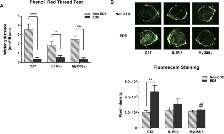
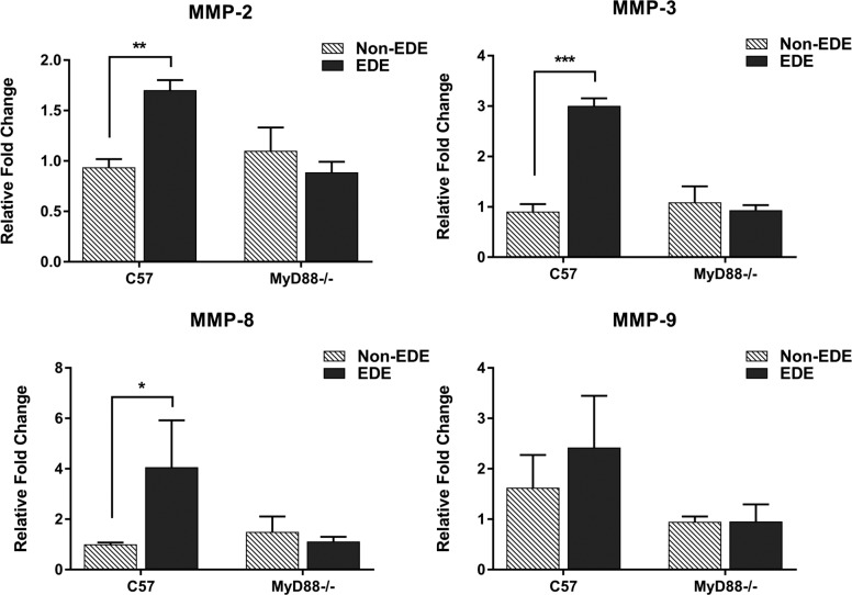
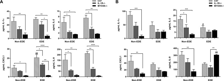
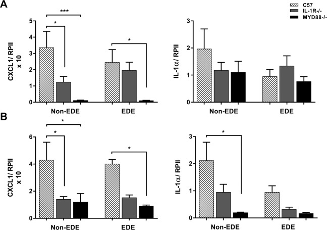
Methods: C57BL/6 wild-type (WT), MyD88-/-, and IL-1R-/- mice were exposed to EDE conditions for 5 days. Tear production was measured by phenol red thread test and ocular surface damage assessed with fluorescein staining. Corneal homogenates were obtained for matrix metalloproteinase (MMP) and cytokine expression analysis by Luminex assay and quantitative PCR. In addition, whole eyes and eyelids were dissected and goblet cells and Meibomian glands were imaged, respectively.

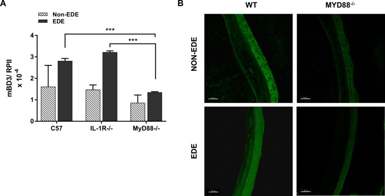
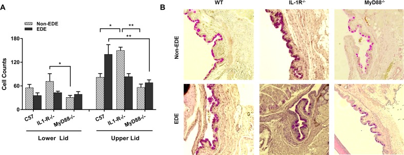
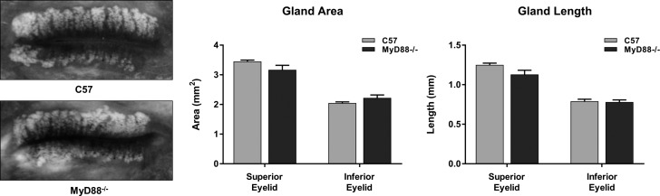
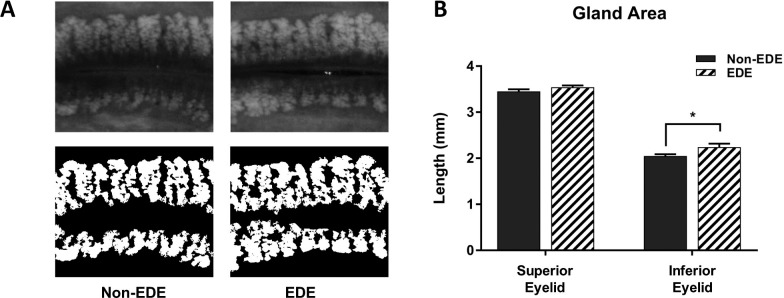
Results: Following 5 days of EDE, WT mice had extensive ocular surface staining, while MyD88-/- mice had no increased staining above non-EDE conditions. Similarly, MyD88-/- mice did not have increased corneal MMP-2, 3, or 8 concentrations, as seen with WT mice. MyD88-deficiency also resulted in decreased corneal cytokine levels. In addition, MyD88-/- mice had significantly lower conjunctival goblet cell counts compared with both WT (EDE) and IL-1R-/- (non-EDE) mice. However, there was no difference in Meibomian gland morphology between WT, IL-1R-/-, and MyD88-/- mice.

Conclusions: These studies demonstrate the importance of TLR signaling in dry eye development. Mice lacking TLR signaling, MyD88-/-, were protected from EDE-induced ocular surface damage and inflammatory mediator expression, warranting further investigation into TLR inhibition as a potential therapeutic for DED.


Figures

Figure 1
Figure 1
EDE results in decreased tear production; however, MyD88−/− mice are protected from ocular surface damage. Mice from each genotype were housed in a control environment (non-EDE) or exposed to desiccating EDE conditions for 5 days (EDE). (A) Tear volume was measured by phenol red thread test. Wicking distance of tears was measured over 15 seconds and length measured in millimeters. Data represent mean ± SEM of six mice per group with analysis by ANOVA with Bonferroni's test for multiple comparisons, *P < 0.05, ***P < 0.001, ****P < 0.0001. (B) Corneal fluorescein staining was performed to examine surface defects using SD-OCT. Images are representative of six mice per group. Data represent mean ± SEM with analysis by ANOVA and Bonferroni's test for multiple comparisons, **P < 0.01, ##P < 0.01 in comparison to C57 EDE.
Figure 2
Figure 2
EDE conditions increase relative MMP expression in WT (C57) but not MyD88−/− corneas. MMP protein expression was determined in WT (C57) and MyD88−/− corneal cell lysates in either non-EDE or EDE conditions by Luminex multiplex assay. Three to five mice were pooled for each sample. Graphs represent mean ± SEM of three independent experiments (n = 3) and show the relative fold change of MMP expression following EDE compared with non-EDE levels. Statistical analysis was by unpaired Student's t-test, *P < 0.05, **P < 0.01, ***P < 0.001.
Figure 3
Figure 3
Ocular surface cytokine protein expression is decreased in MyD88−/− animals during EDE conditions. IL-1α, CXCL1, IL-9, and IL-2 expression were quantitated in corneal and conjunctival homogenates from WT (C57) and MyD88−/− mice after 5 days of EDE treatment or normal (non-EDE) conditions by Luminex multiplex assay. Data represent mean ± SEM of three independent experiments with each sample pooled from five mice. Analysis was by ANOVA with Bonferroni's test for multiple comparisons, with comparison to C57 samples, **P < 0.01, ***P < 0.001.
Figure 4
Figure 4
MyD88-deficient mice have lower cytokine, chemokine, and antimicrobial peptide expression following EDE. Following EDE or normal (non-EDE) conditions, (A) IL-1α, IL-1β, TNFα, and CXCL1 expression were determined in corneal and conjunctival lysates of WT (C57), IL-1R−/−, and MyD88−/− mice by qPCR. Graphs represent mean ± SEM of three independent experiments with each sample pooled from three to four mice. Analysis was by (A) ANOVA with Bonferroni's test for multiple comparisons, *P < 0.05, ****P < 0.0001 and (B) Student's t-test, **P < 0.01.
Figure 5
Figure 5
MyD88−/− mice have decreased expression of the antimicrobial peptide mBD3 following EDE. (A) Following EDE, mBD3 expression was determined in corneal lysates of WT (C57) and MyD88−/− mice by qPCR. Graph represents mean ± SEM of three independent experiments with each sample pooled from three to four mice. Analysis was by ANOVA with Bonferroni's test for multiple comparisons, ***P < 0.001. (B) Frozen corneal sections were stained and imaged for mBD3 expression in non-EDE and EDE conditions. Images are representative of two independent experiments. Scale bars: 50 μm.
Figure 6
Figure 6
MyD88-deficient mice have lower conjunctival goblet cell counts. Paraffin-embedded sections from WT (C57), IL-1R−/−, and MyD88−/− mice were stained with PAS and hematoxylin to visualize goblet cells in the lower and upper conjunctival lid regions. (A) Cells were manually counted and averaged from three to four eyelids per genotype and condition (bottom panel). Graph represents mean ± SEM with analysis by ANOVA and Bonferroni's test for multiple comparisons, *P < 0.05, **P < 0.01, ****P < 0.0001; #P < 0.0001 compared with C57. (B) Representative images of goblet cell PAS staining.
Figure 7
Figure 7
Meibomian gland area and length do not differ between C57 and MyD88−/− mice. The superior and inferior eyelids from WT (C57) and MyD88−/− mice were isolated and imaged with a Keratograph 5M infrared camera. A region of interest from the center of the eyelid containing the superior (upper) and inferior (lower) Meibomian glands was selected and determination of area (mm2) and length (mm) was performed using a custom designed MATLAB software program. Graph represents mean ± SEM with analysis by ANOVA and Bonferroni's test for multiple comparisons, with no significance found between C57 and MyD88−/− mice.
Figure 8
Figure 8
Meibomian gland area is enlarged following EDE. Following 5 days of EDE, the superior and inferior eyelids from 12-week old mice were isolated and imaged with a Keratograph 5M infrared camera. (A) A region of interest from the center of each eyelid was selected (upper panels) and determination of area (mm2) was performed using a custom designed MATLAB software program (bottom panels). (B) Graphs represent mean ± SEM with analysis by two-tailed Student's t-test, *P < 0.05.

References

    1. Buchholz P, Steeds CS, Stern LS,et al. . Utility assessment to measure the impact of dry eye disease. Ocul Surf. 2006; 4: 155– 161. - PubMed
    1. Balik J. . The lacrimal fluid in keratoconjunctivitis sicca; a quantitative and qualitative investigation. Am J Ophthalmol. 1952; 35: 1773– 1782. - PubMed
    1. Pflugfelder SC, Jones D, Ji Z, Afonso A, Monroy D. . Altered cytokine balance in the tear fluid and conjunctiva of patients with Sjögren's syndrome keratoconjunctivitis sicca. Curr Eye Res. 1999; 19: 201– 211. - PubMed
    1. Solomon A, Dursun D, Liu Z, Xie Y, Macri A, Pflugfelder SC. . Pro- and anti-inflammatory forms of interleukin-1 in the tear fluid and conjunctiva of patients with dry-eye disease. Invest Ophthalmol Vis Sci. 2001; 42: 2283– 2292. - PubMed
    1. Chauhan SK, Dana R. . Role of Th17 cells in the immunopathogenesis of dry eye disease. Mucosal Immunol. 2009; 2: 375– 376. - PMC - PubMed

Publication types

MeSH terms

Supplementary concepts

LinkOut - more resources