

Fluorescence Lifetime Imaging Ophthalmoscopy (FLIO) of Macular Pigment
- PMID: 30025128
- PMCID: PMC6009392
- DOI: 10.1167/iovs.18-23886
Fluorescence Lifetime Imaging Ophthalmoscopy (FLIO) of Macular Pigment
Abstract
Purpose: To describe different patterns of macular pigment (MP) seen in fluorescence lifetime imaging ophthalmoscopy (FLIO) and to analyze ex vivo fluorescence characteristics of carotenoids.
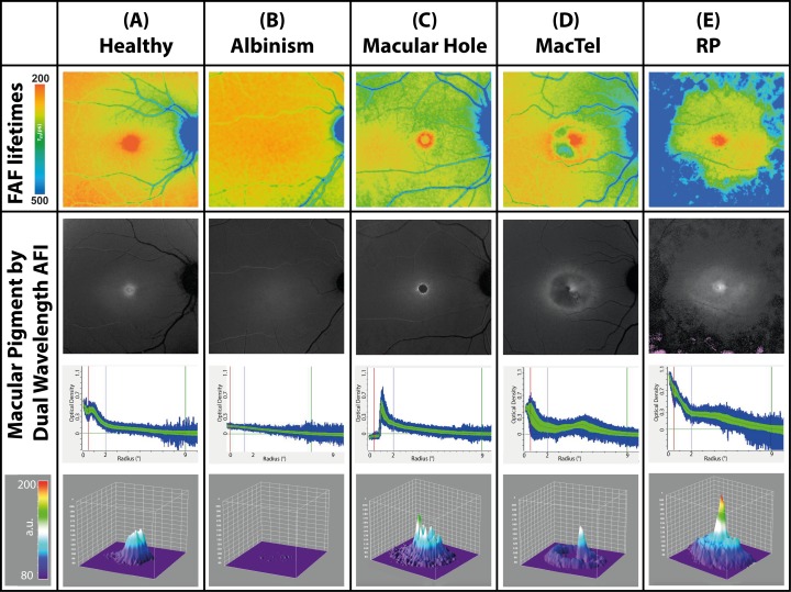
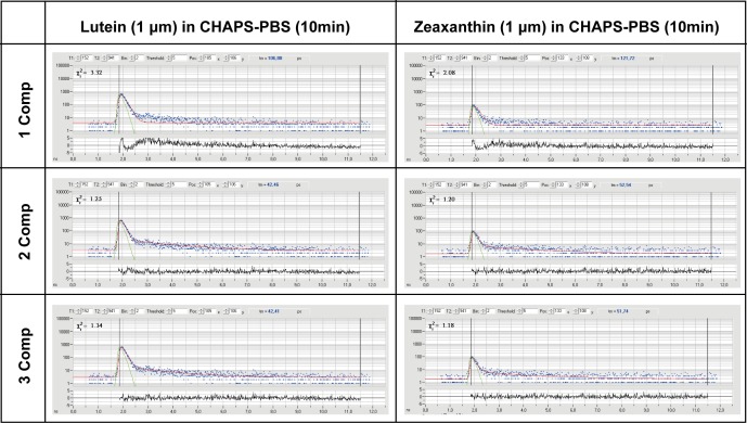
Methods: A total of 31 eyes of young healthy subjects, 4 eyes from patients with albinism, 36 eyes with macular telangiectasia type 2 (MacTel), 24 eyes with retinitis pigmentosa, and 1 eye with a macular hole were included in this clinic-based, cross-sectional study. All subjects underwent Heidelberg Engineering FLIO and MP measurements (dual-wavelength autofluorescence). Fundus autofluorescence (FAF) lifetimes of a 30° retinal field were detected in two spectral channels (SSC: 498-560 nm; LSC: 560-720 nm), and amplitude-weighted mean fluorescence lifetimes (τm) were calculated. Additionally, autofluorescence lifetimes of known dilutions of lutein and zeaxanthin were measured in a cuvette in free- and protein-associated states.
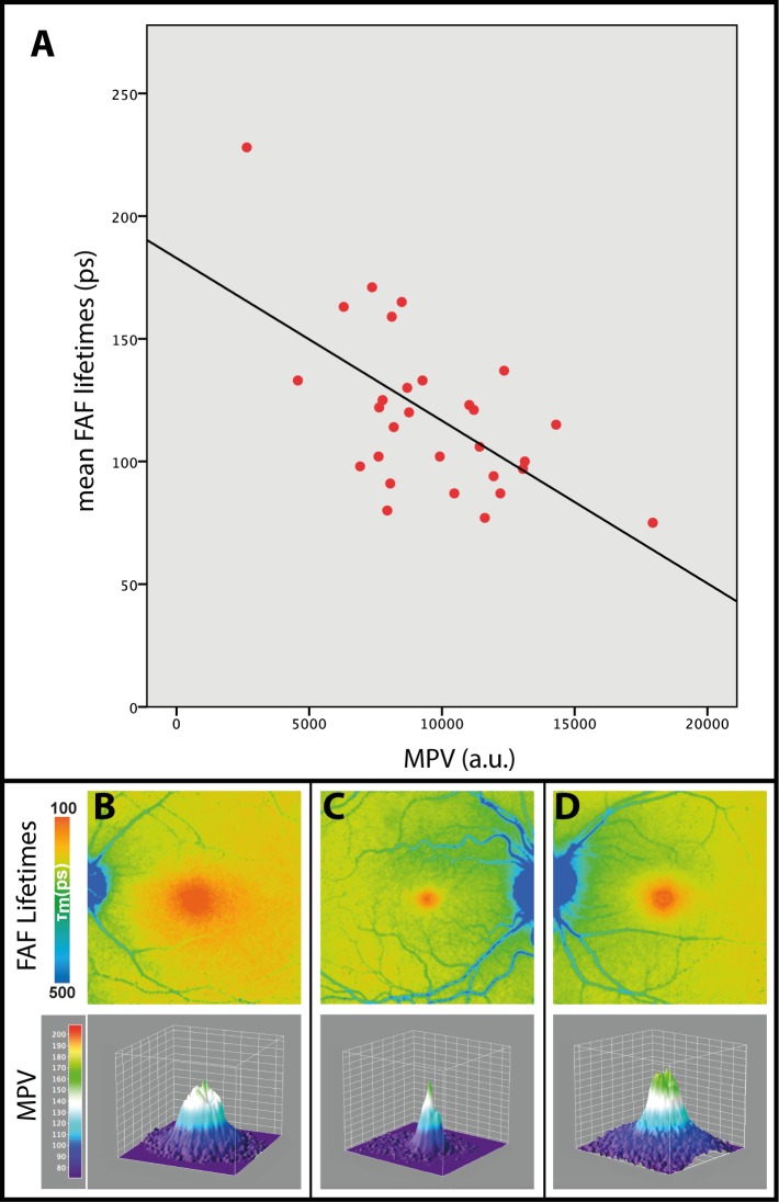
Results: MP shows a significant inverse correlation to foveal FAF lifetimes measured with FLIO (SSC: r = -0.608; P < 0.001). Different distribution patterns can be assigned to specific disease-related changes. Two patients with albinism, who did not have MP, were found to be missing short FAF lifetimes. In solvent, lutein and zeaxanthin show very short autofluorescence lifetimes (∼50-60 ps; SSC), as do their respective binding proteins (∼40-50 ps; SSC). When combining carotenoids with their specific binding proteins, the decay times shift to longer means (∼70-90 ps; SSC).
Conclusions: This study expands upon previous findings of an impact of MP on short FAF lifetimes by describing ex vivo autofluorescence lifetimes of carotenoids and different in vivo autofluorescence patterns that can be associated with certain diseases.
Figures
References
-
- Schweitzer D, Schenke S, Hammer M,et al. . Toward metabolic mapping of the human retina. Microsc Res Tech. 2007; 70: 410– 419. - PubMed
-
- Lakowicz JR. . Principles of Fluorescence Spectroscopy: Springer; 2007.
-
- Becker W. . Fluorescence lifetime imaging--techniques and applications. J Microsc. 2012; 247: 119– 136. - PubMed
Publication types
MeSH terms
Substances
Grants and funding
LinkOut - more resources
Full Text Sources
Other Literature Sources
