. 2018 Sep;38(9):2016-2027.
doi: 10.1161/ATVBAHA.118.311266.

Transcription Factor Zhx2 Deficiency Reduces Atherosclerosis and Promotes Macrophage Apoptosis in Mice

Affiliations

Transcription Factor Zhx2 Deficiency Reduces Atherosclerosis and Promotes Macrophage Apoptosis in Mice

Ayca Erbilgin et al. Arterioscler Thromb Vasc Biol. 2018 Sep.

Abstract

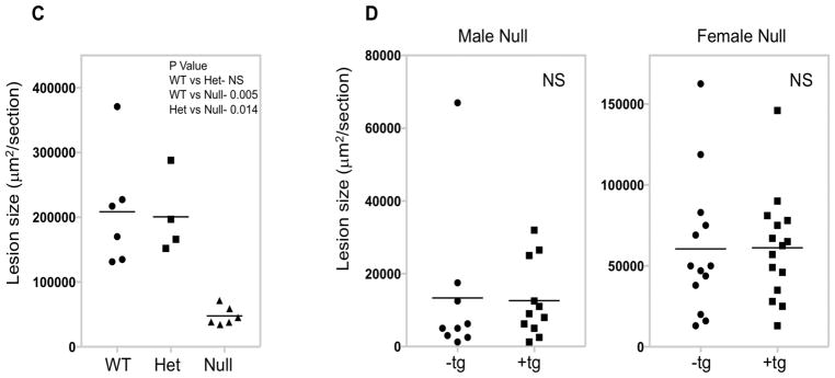
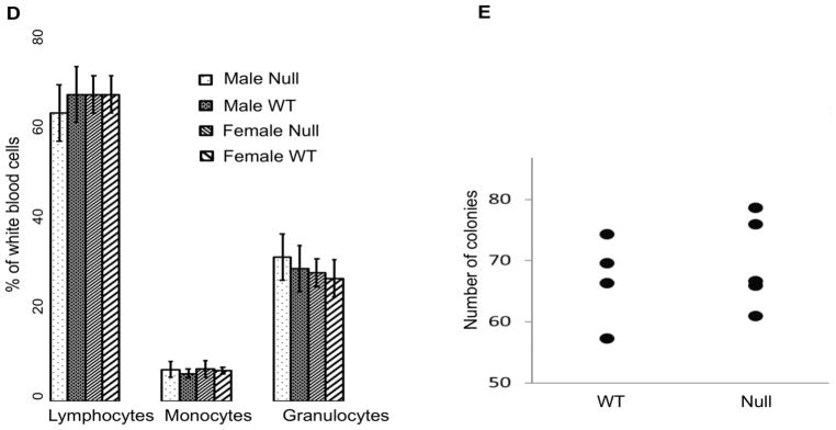
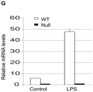
Objective- The objective of this study was to determine the basis of resistance to atherosclerosis of inbred mouse strain BALB/cJ. Approach and Results- BALB/cJ mice carry a naturally occurring null mutation of the gene encoding the transcription factor Zhx2, and genetic analyses suggested that this may confer resistance to atherosclerosis. On a hyperlipidemic low-density lipoprotein receptor null background, BALB/cJ mice carrying the mutant allele for Zhx2 exhibited up to a 10-fold reduction in lesion size as compared with an isogenic strain carrying the wild-type allele. Several lines of evidence, including bone marrow transplantation studies, indicate that this effect of Zhx2 is mediated, in part, by monocytes/macrophages although nonbone marrow-derived pathways are clearly involved as well. Both in culture and in atherosclerotic lesions, macrophages from Zhx2 null mice exhibited substantially increased apoptosis. Zhx2 null macrophages were also enriched for M2 markers. Effects of Zhx2 on proliferation and other bone marrow-derived cells, such as lymphocytes, were at most modest. Expression microarray analyses identified >1000 differentially expressed transcripts between Zhx2 wild-type and null macrophages. To identify the global targets of Zhx2, we performed ChIP-seq (chromatin immunoprecipitation sequencing) studies with the macrophage cell line RAW264.7. The ChIP-seq peaks overlapped significantly with gene expression and together suggested roles for transcriptional repression and apoptosis. Conclusions- A mutation of Zhx2 carried in BALB/cJ mice is responsible in large part for its relative resistance to atherosclerosis. Our results indicate that Zhx2 promotes macrophage survival and proinflammatory functions in atherosclerotic lesions, thereby contributing to lesion growth.

Keywords: apoptosis; atherosclerosis; bone marrow; genetics; macrophages; mice; transcription factor.


Figures

Figure 1
Figure 1. Zhx2 deficiency suppresses atherosclerosis in BALB/cJ Ldlr−/− mice independently of expression in liver or effects on plasma lipids
(A) Ldlr−/− Zhx2 wt and Zhx2 null mice on a BALB/cJ Ldlr−/− background were maintained on a Western diet for 18 weeks and atherosclerotic lesions were quantitated. Aortic root measurements for male and female mice of both genotypes are shown (n=7–15). (B) En face atherosclerosis measurements for male Zhx2 wt (n=7) and Zhx2 null (n=4) mice on a BALB/cJ, Ldlr−/− background. (C) Male BALB/cJ Ldlr−/− mice of Zhx2 genotypes +/+, +/−, −/− were maintained on a Western diet for 18 weeks and lesions were quantitated (± SEM). (D) Atherosclerosis in male Ldlr−/−, Zhx2 null mice is not significantly altered by transgenic (Tg) expression of Zhx2 in liver using a transthyretin (TTR) promoter.
Figure 2
Figure 2. The effects of Zhx2 on atherosclerosis are mediated in part by bone marrow-derived cells
(A): Representative images of immunostaining of lesion macrophages. Red: Mac3; Blue, DAPI. Original magnification, 10× (B): Aortic root lesions of BALB/cJ Ldlr−/− Zhx2 wt or Zhx2 null bone marrow transplants into Ldlr−/− Zhx2 wt (C): Aortic root lesions of Ldlr−/− Zhx2 null bone marrow transplants into Ldlr−/− Zhx2 wt or Zhx2 null recipients (D): Circulating leukocyte levels in Zhx2 null and wt mice on a Western diet were examined using a HemaTrue analyzer. Shown is a representative experiment (N=7–11 mice per condition, + SD). (E): The number of committed bone marrow stem cells responsive to M-CSF did not differ between Zhx2 null and Zhx2 wt mice.
Figure 3
Figure 3. Zhx2 deficiency promotes macrophage apoptosis and an M2 phenotype in vitro and in lesions
(A) Representative sections of lesions from ZhX2 wt (top) and ZhX2 null (bottom) mice stained for TUNEL or CD68. The last panel shows the merged image, magnification X20 (B) Quantitation of TUNEL positive macrophages in lesions (n=4 mice). (C) In vitro TUNEL assay in cultured BMDM from Zhx2 wt and null macrophages with and without oxidized LDL (oxLDL) and 7-ketocholesterol (Keto). (D) Quantitation of TUNEL positive macrophages in vitro with and without oxLDL and Keto (n=3 mice, 4–5 observation from each mice). (E) In vitro apoptosis by TUNEL assay of peritoneal macrophages isolated from the mice maintained on chow or Western diets. (F) Quantitation of TUNEL positive peritoneal macrophages (n= 4 mice, 1–2 observation from each mice) (G) Peritoneal macrophages from BALB/cJ, Ldlr−/− male mice were treated with bacterial lipopolysaccharide (LPS) and Zhx2 transcript levels quantitated by quantitative PCR. (H)Lesional macrophages for Zhx2 wt and null mice were examined for M1 markers using flow cytometry. A significantly higher percentage of cells expressed the M1 marker CD86 (n= 4 mice, aorta from 2 mice were combined in each group).
Figure 4
Figure 4. ChIP-seq analysis of Zhx2 in the RAW macrophage cell line
(A) Examples of enrichment of ChIP-seq peaks at two genes in Jun and Bcl6. (B) Transcription factor motifs enriched in Zhx2 ChIP-seq peaks, including p-values and best match details.

References

    1. Gu L, Johnson MW, Lusis AJ. Quantitative trait locus analysis of plasma lipoprotein levels in an autoimmune mouse model : interactions between lipoprotein metabolism, autoimmune disease, and atherogenesis. Arterioscler Thromb Vasc Biol. 1999;19:442–453. - PubMed
    1. Gargalovic PS, Erbilgin A, Kohannim O, Pagnon J, Wang X, Castellani L, LeBoeuf R, Peterson ML, Spear BT, Lusis AJ. Quantitative trait locus mapping and identification of Zhx2 as a novel regulator of plasma lipid metabolism. Circ Cardiovasc Genet. 2010;3:60–67. - PMC - PubMed
    1. Wang X, Gargalovic P, Wong J, Gu JL, Wu X, Qi H, Wen P, Xi L, Tan B, Gogliotti R, Castellani LW, Chatterjee A, Lusis AJ. Hyplip2, a new gene for combined hyperlipidemia and increased atherosclerosis. Arterioscler Thromb Vasc Biol. 2004;24:1928–1934. - PubMed
    1. Perincheri S, Dingle RW, Peterson ML, Spear BT. Hereditary persistence of alpha-fetoprotein and H19 expression in liver of BALB/cJ mice is due to a retrovirus insertion in the Zhx2 gene. Proc Natl Acad Sci U S A. 2005;102:396–401. - PMC - PubMed
    1. Yue X, Zhang Z, Liang X, Gao L, Zhang X, Zhao D, Liu X, Ma H, Guo M, Spear BT, Gong Y, Ma C. Zinc fingers and homeoboxes 2 inhibits hepatocellular carcinoma cell proliferation and represses expression of Cyclins A and E. Gastroenterology. 2012;142:1559–1570. - PMC - PubMed

Publication types