. 2018 Jul 20;9(7):735.
doi: 10.1038/s41419-018-0765-9.

Reversing wrinkled skin and hair loss in mice by restoring mitochondrial function

Affiliations

Reversing wrinkled skin and hair loss in mice by restoring mitochondrial function

Bhupendra Singh et al. Cell Death Dis. .

Abstract

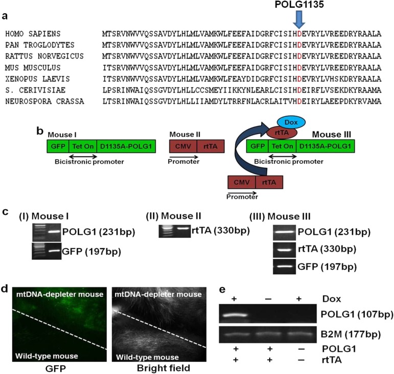
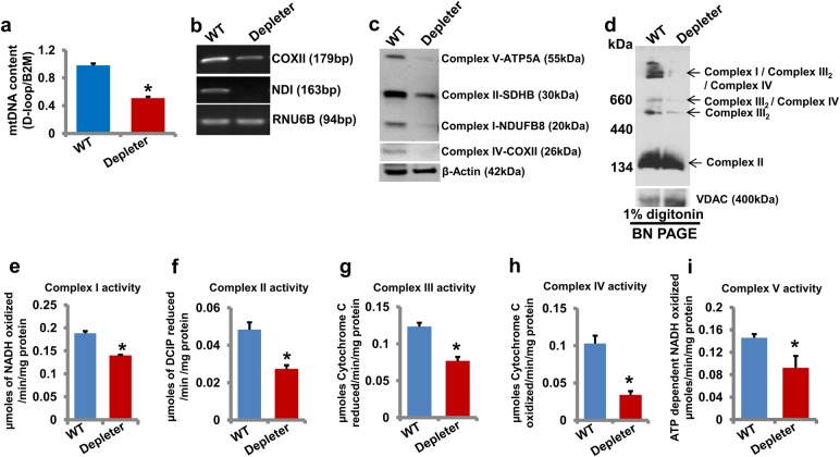
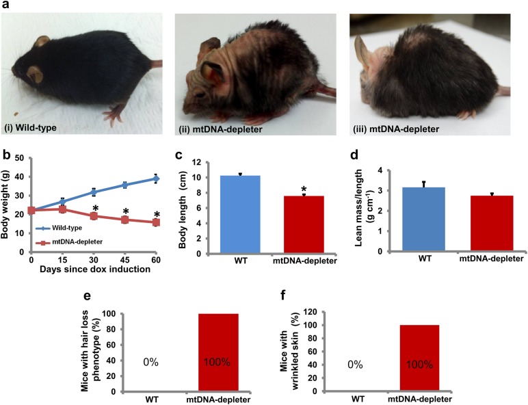
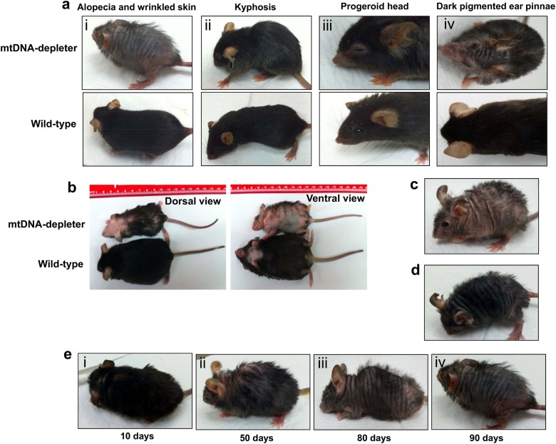
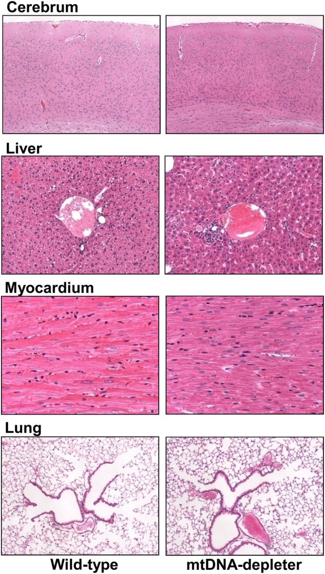
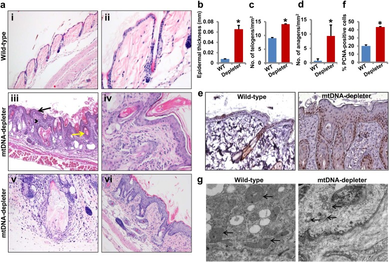
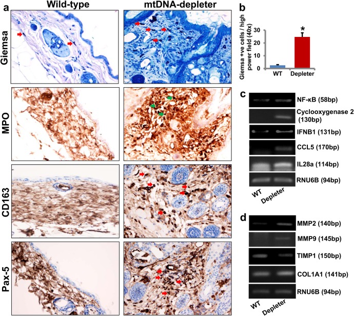
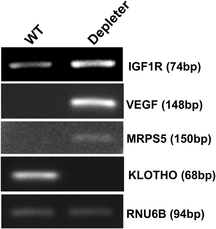
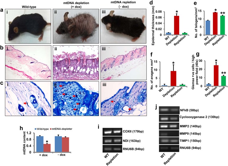
Mitochondrial DNA (mtDNA) depletion is involved in mtDNA depletion syndromes, mitochondrial diseases, aging and aging-associated chronic diseases, and other human pathologies. To evaluate the consequences of depletion of mtDNA in the whole animal, we created an inducible mtDNA-depleter mouse expressing, in the polymerase domain of POLG1, a dominant-negative mutation to induce depletion of mtDNA in various tissues. These mice showed reduced mtDNA content, reduced mitochondrial gene expression, and instability of supercomplexes involved in oxidative phosphorylation (OXPHOS) resulting in reduced OXPHOS enzymatic activities. We demonstrate that ubiquitous depletion of mtDNA in mice leads to predominant and profound effects on the skin resulting in wrinkles and visual hair loss with an increased number of dysfunctional hair follicles and inflammatory responses. Development of skin wrinkle was associated with the significant epidermal hyperplasia, hyperkeratosis, increased expression of matrix metalloproteinases, and decreased expression of matrix metalloproteinase inhibitor TIMP1. We also discovered markedly increased skin inflammation that appears to be a contributing factor in skin pathology. Histopathologic analyses revealed dysfunctional hair follicles. mtDNA-depleter mice also show changes in expression of aging-associated markers including IGF1R, KLOTHO, VEGF, and MRPS5. mtDNA-repleter mice showed that, by turning off the mutant POLG1 transgene expression, mitochondrial function, as well as the skin and hair pathology, is reversed to wild-type level. To our knowledge that restoration of mitochondrial functions can reverse the skin and hair pathology is unprecedented.


Conflict of interest statement

The authors declare that they have no conflict of interest.

Figures

Fig. 1
Fig. 1. Creation and verification of doxycycline-inducible mtDNA-depleter mice
a Alignment of amino acid sequences of polymerase domain of POLG1 protein from Homo sapiens to Neurospora crassa shows that aspartic acid in the POLG1 at 1135 position is evolutionarily conserved. b Schematic of the development of inducible D1135A-POLG1 (mtDNA-depleter) transgenic mouse model. D1135A-POLG1-expressing mouse (Mouse I) was created by microinjection of the pTRE-Tight-BI-AcGFP1-D1135-POLG1 construct into the one-cell stage egg from C57BL/6 mouse. The D1135A-POLG1-positive founder male mouse (Mouse I) was bred with the CAG-rtTA3 female mouse (Mouse II, Jackson Laboratories, stock # 016532) to get the D1135A-POLG1 transgenic animal (Mouse III). c Pups genotyping reveals the presence of D1135A-POLG1, rtTA, and GFP. d Whole-body imaging also confirms expression of GFP in only mtDNA-depleter mice. e RT-PCR analyses confirm dox-dependent expression of D1135A-POLG1 in only mtDNA-depleter mice
Fig. 2
Fig. 2. Analyses of mtDNA content, gene expression, and OXPHOS activity in mtDNA-depleter mice
a Quantification of mtDNA content (mean ± s.e.m; *P<0.05, Student’s t test) in skin samples from wild-type control (WT; n = 3) and mtDNA-depleter (Depleter; n = 3) mice after 2 months of continuous dox induction. bd RT-PCR analysis of mtDNA-encoded genes (b), Western blot analysis of OXPHOS subunits (c), and BN-PAGE analysis of OXPHOS supercomplexes (d) in the skin of wild-type control and mtDNA-depleter mice after 2 months of continuous dox induction. e–i Enzymatic activities of OXPHOS complex I (e), II (f), III (g), IV (h), and V (i) in the skin of wild-type control and mtDNA-depleter mice after 2 months of continuous dox induction. Depleter = mtDNA-depleter mice
Fig. 3
Fig. 3. Skin wrinkles and hair loss in mtDNA-depleter mice
a mtDNA-depleter mice show skin wrinkles (ii), hair loss (ii), and kyphosis (iii) after 4–8 weeks of continuous dox-mediated induction. bd Quantitative assessment of body weight (b), body length (c), and lean mass/length ratio (d) of mtDNA-depleter (n = 30) and wild-type control mice (n = 30). Data are expressed as mean ± s.e.m; *P<0.05, Student’s t test. e, f Quantitative assessment of hair loss (e) and wrinkled skin (f) phenotypic changes in mtDNA-depleter (n = 30) and wild-type control mice (n = 30) after 60 days of continuous dox induction
Fig. 4
Fig. 4. Additional phenotypic changes in mtDNA-depleter mice
a mtDNA-depleter mice demonstrate a very strong alopecia and wrinkled skin (i), kyphosis (ii), progeroid head (iii), and darkly pigmented ear pinnae (iv) phenotypic changes after induction with dox. b Representative images of a mtDNA-depleter mouse showing the gross phenotypic changes in the size and appearance compared to age-matched wild-type control littermate. c, d The different patterns of hair loss in male (c) and female (d) mtDNA-depleter mice. e Representative images showing gradual time-dependent phenotypic changes in skin wrinkles and hair loss in a female mtDNA-depleter mouse after continuous dox induction (i–iv)
Fig. 5
Fig. 5. Histological analyses of different tissues from mtDNA-depleter mice
Representative hematoxylin- and eosin-stained cross-sections of brain (cerebrum), liver, heart (myocardium), and lung from wild-type control (n = 3) and mtDNA-depleter mice (n = 3) after 2 months of dox induction
Fig. 6
Fig. 6. Histological and microscopic analyses of skin of mtDNA-depleter mice
a Hematoxylin- and eosin-stained sections of dorsal skin from wild-type control (n = 3) (i and ii) and mtDNA-depleter mice (n = 3) (iii–-vi) after 2 months of continuous dox induction. While the skin of wild-type mice shows the presence of normal skin histology (i, ×10), the skin of mtDNA-depleter mice shows hyperplastic epidermis with hyperkeratosis (black color arrow), dysfunctional hair follicles containing keratinaceous debris and/or malformed hair (yellow color arrow), and increased the number of inflammatory cells in the dermis (arrowhead) (iii, ×10). Skin sections at higher magnification show the presence of normal telogen hair follicles (ii, ×40) in wild-type control mice and aberrant telogen (iv, ×40) and anagen hair follicles (vi, ×20) with defective sebaceous glands. Panel v shows ruptured follicular cyst surrounded by granulomatous and mixed inflammatory infiltrate in mtDNA-depleter mice. bd Quantification of epidermal thickness (b), hair follicles in telogen (c), and anagen (d) stages of hair cycle (mean ± s.e.m; *P<0.05, Student’s t test) in skin samples from wild-type control (n = 3) and mtDNA-depleter (n = 3) mice after 2 months of continuous dox induction. e Representative images of PCNA immuno-stained cross-sections of skin from wild-type control (n = 3) and mtDNA-depleter mice (n = 3) after 2 months of dox induction. The basement membrane position in these images is marked with dotted lines. f Quantification of epidermal proliferation (PCNA+) in skin samples from wild-type control (n = 3) and mtDNA-depleter (n = 3) mice after 2 months of continuous dox induction. g Electron micrographs of skin samples from wild-type control (n = 3) and mtDNA-depleter mice (n = 3) after 2 months of dox induction. Skin from mtDNA-depleter mice revealed a severely disturbed mitochondrial structure with loss of cristae and degeneration of intramitochondrial structures. Depleter = mtDNA-depleter mice
Fig. 7
Fig. 7. Skin inflammation in mtDNA-depleter mice
a Immunocytochemical and histochemical analyses of skin sections show the presence of increased number of inflammatory cells including mast cells (Giemsa stain-positive cells), granulocytes (MPO-positive cells), macrophages and histiocytes (CD163-positive cells), and B lymphocytes (Pax-5-positive cells) in the dermis, as well as in perifollicular and periepidermal location of mtDNA-depleter mice. The skin sections of wild-type mice are predominantly negative for MPO, CD163, and Pax-5 staining. Arrows indicate the presence of inflammatory cells in the skin sections. b Quantitative analysis of Giemsa-positive mast cells in the skin sections of wild-type control and mtDNA-depleter mice (mean ± s.e.m; *P<0.05, Student’s t test). c RT-PCR analysis of inflammatory genes in the skin RNA samples of wild-type control (WT; n = 3) and mtDNA-depleter mice (Depleter; n = 3) after 2 months of continuous dox induction. d RT-PCR analysis of genes in the skin RNA samples of wild-type control (n = 3) and mtDNA-depleter mice (n = 3) after 2 months of continuous dox induction. Depleter = mtDNA-depleter mice
Fig. 8
Fig. 8. Expression of aging-associated markers in mtDNA-depleter mice
Representative images showing mRNA expression analyses of IGF1R, VEGF, MRPS5, and Klotho genes (marker genes of intrinsic aging) by RT-PCR in the skin samples of wild-type control (n = 3) and mtDNA-depleter mice (n = 3) after 2 months of dox induction. Depleter = mtDNA-depleter mice
Fig. 9
Fig. 9. Reversal of wrinkled skin and hair loss to wild-type by restoration of mitochondrial function.
a Representative images of a mtDNA-depleter mouse showing skin wrinkles and hair loss after 2 months of continuous dox induction (+dox; mtDNA depletion) (ii) and reversal of these phenotypic changes after 1 month of dox withdrawal (−dox; mtDNA repletion) (iii; n= 3). Wild-type control mice (n = 3) did not show any change in skin phenotype after dox treatment or after 1 month of dox withdrawal (i). b Hematoxylin- and eosin-stained sections of dorsal skin from wild-type control mice (i), mtDNA-depleter mice after 2 months of continuous dox induction (mtDNA depletion) (ii) and after 1 month of dox withdrawal (mtDNA repletion) (iii). c Giemsa staining of skin sections shows the presence of increased number of mast cells in the dermis and perifollicular as well as near-epidermal locations of mtDNA-depleter mice compared with skin sections of wild-type mice. Compared with mtDNA-depleter mice, the lower number of mast cells is present in the skin of mtDNA-depleter mice after 1 month of dox withdrawal (−dox; mtDNA repletion). Arrows indicate the presence of Giemsa-positive cells in the skin sections. df Quantification of epidermal thickness (d), hair follicles in telogen (e), and anagen (f) stages of hair cycles (mean ± s.e.m; *P<0.05, Student’s t test) in skin samples from wild-type control (n = 3) and mtDNA-depleter (n = 3) mice after 2 months of continuous dox induction (mtDNA depletion) and after 1 month of dox withdrawal (mtDNA repletion). g Quantitative analysis of Giemsa-positive mast cells in the skin sections of wild-type control, mtDNA-depleter mice after 2 months of continuous dox induction (mtDNA depletion), and after 1 month of dox withdrawal (mtDNA repletion) (mean ± s.e.m; *P<0.05, Student’s t test). h Quantification of mtDNA content (mean ± s.e.m; *P<0.05, Student’s t test) in skin samples from wild-type control (n = 3) and mtDNA-depleter (n = 3) mice after 2 months of continuous dox induction and reversal of mtDNA content in skin samples of mtDNA-depleter mice (n = 3) after 1 month of dox withdrawal. i, j Representative gel images showing RT-PCR analysis of genes in the skin RNA samples of wild-type control (n = 3) and mtDNA-depleter mice (n = 3) after 1 month of dox withdrawal (mtDNA repletion). Depletion = mtDNA depletion, repletion = mtDNA repletion

Comment in

  • Mitochondrial DNA keeps you young.
    Bonora M, Pinton P. Bonora M, et al. Cell Death Dis. 2018 Sep 24;9(10):992. doi: 10.1038/s41419-018-1045-4. Cell Death Dis. 2018. PMID: 30250200 Free PMC article. No abstract available.

Similar articles

Cited by

References

    1. Alberio S, Mineri R, Tiranti V, Zeviani M. Depletion of mtDNA: syndromes and genes. Mitochondrion. 2007;7:6–12. doi: 10.1016/j.mito.2006.11.010. - DOI - PubMed
    1. Ryan MT, Hoogenraad NJ. Mitochondrial–nuclear communications. Annu. Rev. Biochem. 2007;76:701–722. doi: 10.1146/annurev.biochem.76.052305.091720. - DOI - PubMed
    1. Tuppen HA, Blakely EL, Turnbull DM, Taylor RW. Mitochondrial DNA mutations and human disease. Biochim. Biophys. Acta. 1797;113-128:2010. - PubMed
    1. El-Hattab AW, Scaglia F. Mitochondrial DNA depletion syndromes: review and updates of genetic basis, manifestations, and therapeutic options. Neurotherapeutics. 2013;10:186–198. doi: 10.1007/s13311-013-0177-6. - DOI - PMC - PubMed
    1. Elpeleg O, et al. Deficiency of the ADP-forming succinyl-CoA synthase activity is associated with encephalomyopathy and mitochondrial DNA depletion. Am. J. Hum. Genet. 2005;76:1081–1086. doi: 10.1086/430843. - DOI - PMC - PubMed

Publication types

MeSH terms