. 2018 Jul 19;8(1):10909.
doi: 10.1038/s41598-018-29101-6.

Small molecule activator of Nm23/NDPK as an inhibitor of metastasis

Affiliations

Small molecule activator of Nm23/NDPK as an inhibitor of metastasis

Jae-Jin Lee et al. Sci Rep. .

Abstract

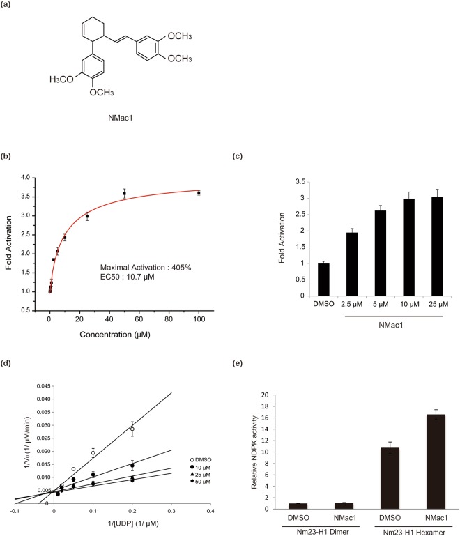
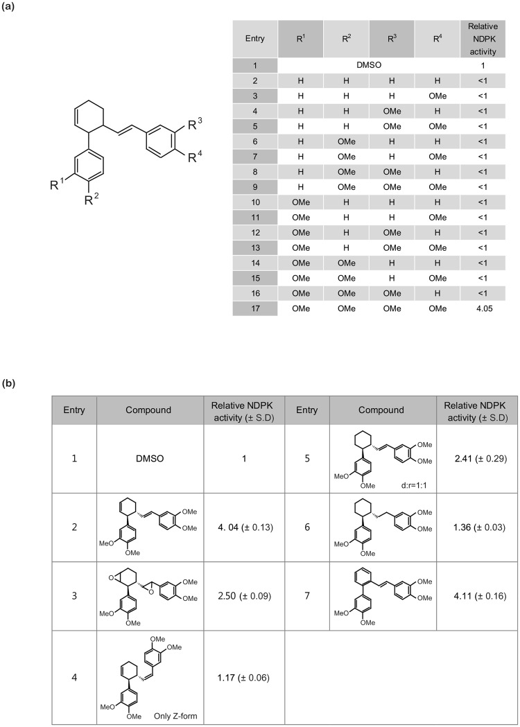
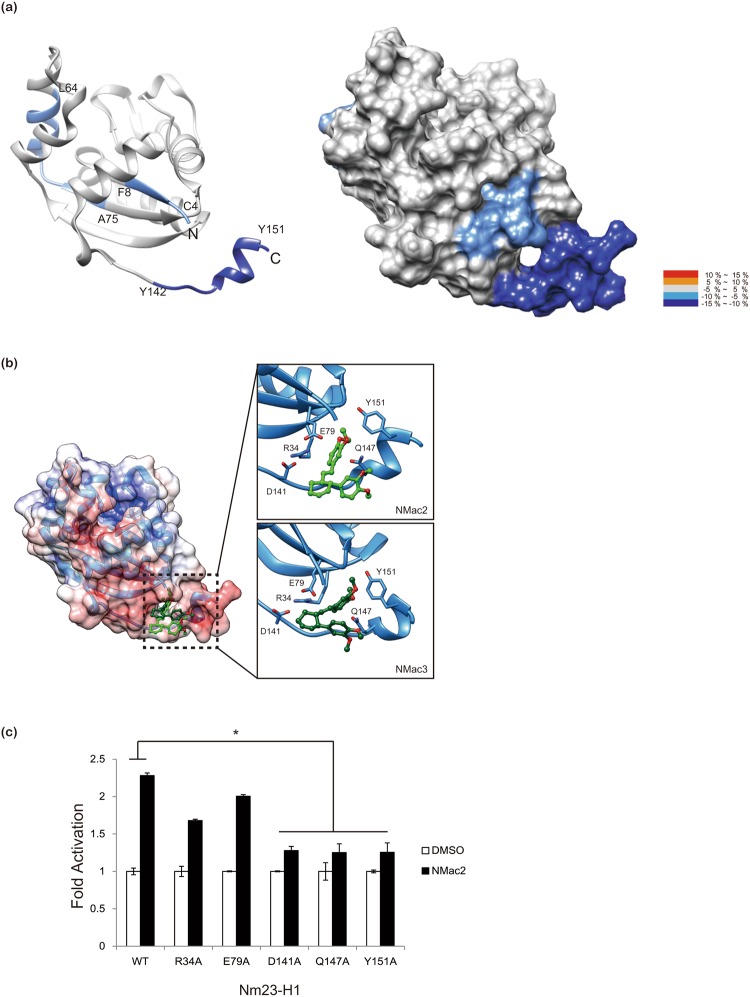
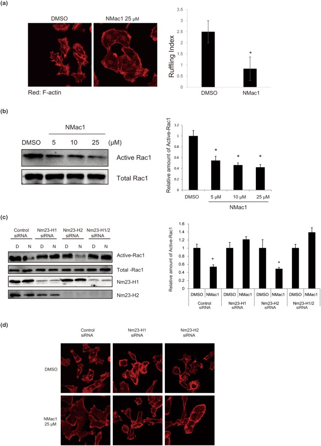
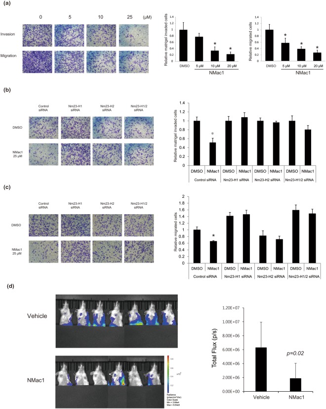
Nm23-H1/NDPK-A is a tumor metastasis suppressor having NDP kinase (NDPK) activity. Nm23-H1 is positively associated with prolonged disease-free survival and good prognosis of cancer patients. Approaches to increasing the cellular levels of Nm23-H1 therefore have significance in the therapy of metastatic cancers. We found a small molecule, (±)-trans-3-(3,4-dimethoxyphenyl)-4-[(E)-3,4-dimethoxystyryl]cyclohex-1-ene, that activates Nm23, hereafter called NMac1. NMac1 directly binds to Nm23-H1 and increases its NDPK activity. Employing various NMac1 derivatives and hydrogen/deuterium mass spectrometry (HDX-MS), we identified the pharmacophore and mode of action of NMac1. We found that NMac1 binds to the C-terminal of Nm23-H1 and induces the NDPK activation through its allosteric conformational changes. NMac1-treated MDA-MB-231 breast cancer cells showed dramatic changes in morphology and actin-cytoskeletal organization following inhibition of Rac1 activation. NMac1 also suppressed invasion and migration in vitro, and metastasis in vivo, in a breast cancer mouse model. NMac1 as an activator of NDPK has potential as an anti-metastatic agent.


Conflict of interest statement

The authors declare no competing interests.

Figures

Figure 1
Figure 1
Identification and characterization of a small molecule NDPK activator of Nm23-H1 and H2. (a) Chemical structure of the NDPK activator, NMac1 (±)-trans-3-(3,4-dimethoxyphenyl)-4-[(E)-3,4-dimethoxystyryl]cyclohex-1-ene. (b) Effect of increasing concentrations of NMac1 on NDPK activity of recombinant human Nm23-H1. NDPK activity was measured by the amount of ATP produced by 5 ng of Nm23-H1 using 5 μM ADP and 5 μM UTP for 1 min with indicated concentration of NMac1, or 1% DMSO as vehicle. All experiments were triplicated, and data are expressed as mean ± S.D. (c) Cell based NDPK assay in a dose dependence. NDPK activity was measured by ATP consumption with 100 μg of MDA-MB-231 cell lysate, and 50 μM UDP. All experiments were triplicated, and data are expressed as mean ± S.D. (d) A double reciprocal plot with fixed concentration of NMac1 and various concentrations of UDP with 5 μM ATP. All experiments were triplicated, and data are expressed as mean ± S.D. (e) NDPK activation by NMac1 to Nm23-H1 hexamer and dimer which were fractionated using size exclusion chromatography. NDPK activity was measured by the amount of ATP produced by 5 ng of Nm23-H1 fraction using 5 μM ADP and 5 μM UTP for 1 min. All experiments were triplicated, and data are expressed as mean ± S.D.
Figure 2
Figure 2
Structure activity relationship (SAR) of NMac1 derivatives for activating NDPK (a) Structure Activity Relationship (SAR) of catechol derivatives. (b) SAR around cyclohexene ring related.
Figure 3
Figure 3
NMac1 binds to C-terminal of Nm23-H1. (a) Overlay of differential HDX data of NMac1 bound to Nm23-H1. This overlay compares HDX of apo Nm23-H1 with that of NMac1 bound Nm23-H1. Perturbation data are color coded and plotted onto the backbone of the 1JXV PDB file according to the key. Observed changes in HDX were statistically significant (p < 0.05) in a two tailed t-test (n = 3) (b) Molecular modeling of interaction between Nm23-H1 and chiral isomer NMac2 and NMac3 (c) Nm23-H1 C-terminal define the allosteric compound binding site. The activity of NMac2 to activate NDPK was examined against a series of Nm23-H1 mutants.
Figure 4
Figure 4
NMac1 induces morphological and other changes in MDA-MB-231 cells. (a) NMac1 reduces membrane ruffle in MDA-MB-231 cells. Localization of F-actin was analyzed by confocal microscopy in control and NMac1 treated cells. MDA-MB-231 cells treated with NMac1 25 µM (or 0.05% DMSO as vehicle) for 16 h were stained by phalloidin-Rhodamine. Number of cell ruffles of MDA-MB-231 cells was quantified by determining the ruffling index. (b) NMac1 reduces Rac1 activation. Active Rac1 pulldown assay was conducted in control and NMac1 treated MDA-MB-231 cells with indicated concentrations for 16 h. A representative experiment out of 3 independent experiments is shown. (c) NMac1 reduces Rac1 activation via Nm23-H1. Active Rac1 pulldown assay was conducted in control and NMac1 treated Nm23-H1 or (/and) H2 knocked down MDA-MB-231 cells with indicated concentrations for 16 h. A representative experiment out of 3 independent experiments is shown. (d) NMac1 reduces membrane ruffle in MDA-MB-231 cell via Nm23-H1. Localization of F-actin was analyzed by confocal microscopy in control and NMac1 treated Nm23-H1 or/and H2 knocking down MDA-MB-231 cells. At 48 hours after Nm23-H1 or/and H2 knocking down, MDA-MB-231 cells were treated with NMac1 25 µM (or 0.05% DMSO) for 16 hours followed by staining with phalloidin-Rhodamine.
Figure 5
Figure 5
Effects of NMac1 on in vitro invasion/migration of MDA-MB-231 cells and in vivo metastasis. (a) NMac1 inhibits transwell migration and matrigel invasion of MDA-MB-231 cells. MDA-MB-231 cells treated with indicated concentrations of NMac1 were allowed to migrate in Boyden chambers for 24 hours. Bars indicate percentage of migration and invasion. *P < 0.05, Student’s t-test. (b) NMac1 inhibits matrigel invasion of MDA-MB-231 cells via Nm23-H1 and H2. Nm23-H1 or (/and) H2 knocking down MDA-MB-231 cells treated with 25 µM of NMac1 were allowed to invaded in matrigel coated Boyden chambers for 24 hours. Bars indicate amount of migration and invasion. *P < 0.05, Student’s t-test. (c) NMac1 inhibits transwell migration of MDA-MB-231 cells via Nm23-H1 and H2. Nm23-H1 or (/and) H2 knocking down MDA-MB-231 cells treated with indicated concentrations of NMac1 were allowed to migrated in Boyden chambers for 24 hours. Bars indicate percentage of migration and invasion. *P < 0.05, Student’s t-test. (d) MDA-MB-231-Luc-D3H2LN cells were orthotopically injected into NOD/SCID mice and the mice were treated with vehicle or NMac1 (n = 7 for VEH group, n = 6 for NMac1 group). (Left) Bioluminescence images visualizing the metastatic tumor cells. (Right) Quantification of bioluminescence images 3 weeks after the NMac1 treatment. Data are presented as mean total photon flux per second ± S.D.

References

    1. Wallet V, et al. Dictyostelium nucleoside diphosphate kinase highly homologous to Nm23 and Awd proteins involved in mammalian tumor metastasis and Drosophila development. J Natl Cancer Inst. 1990;82(14):1199–1202. doi: 10.1093/jnci/82.14.1199. - DOI - PubMed
    1. de la Rosa A, Williams RL, Steeg PS. Nm23/nucleoside diphosphate kinase: toward a structural and biochemical understanding of its biological functions. Bioessays. 1995;17(1):53–62. doi: 10.1002/bies.950170111. - DOI - PubMed
    1. Steeg PS, et al. Evidence for a novel gene associated with low tumor metastatic potential. J Natl Cancer Inst. 1988;80(3):200–204. doi: 10.1093/jnci/80.3.200. - DOI - PubMed
    1. Lacombe ML, Milon L, Munier A, Mehus JG, Lambeth DO. The human Nm23/nucleoside diphosphate kinases. J Bioenerg Biomembr. 2000;32(3):247–258. doi: 10.1023/A:1005584929050. - DOI - PubMed
    1. Ma D, Luyten GP, Luider TM, Jager MJ, Niederkorn JY. Association between NM23-H1 gene expression and metastasis of human uveal melanoma in an animal model. Invest Ophthalmol Vis Sci. 1996;37(11):2293–2301. - PubMed

Publication types

MeSH terms