. 2018 Dec;43(13):2586-2596.
doi: 10.1038/s41386-018-0141-6. Epub 2018 Jul 9.

Neuropsychopharmacology of JNJ-55308942: evaluation of a clinical candidate targeting P2X7 ion channels in animal models of neuroinflammation and anhedonia

Affiliations

Neuropsychopharmacology of JNJ-55308942: evaluation of a clinical candidate targeting P2X7 ion channels in animal models of neuroinflammation and anhedonia

Anindya Bhattacharya et al. Neuropsychopharmacology. 2018 Dec.

Abstract

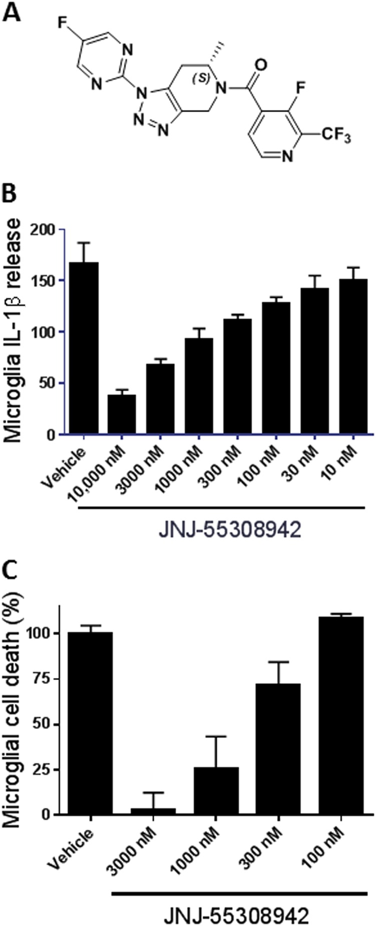
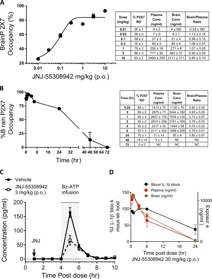
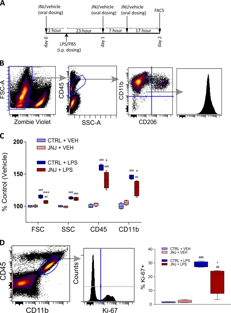
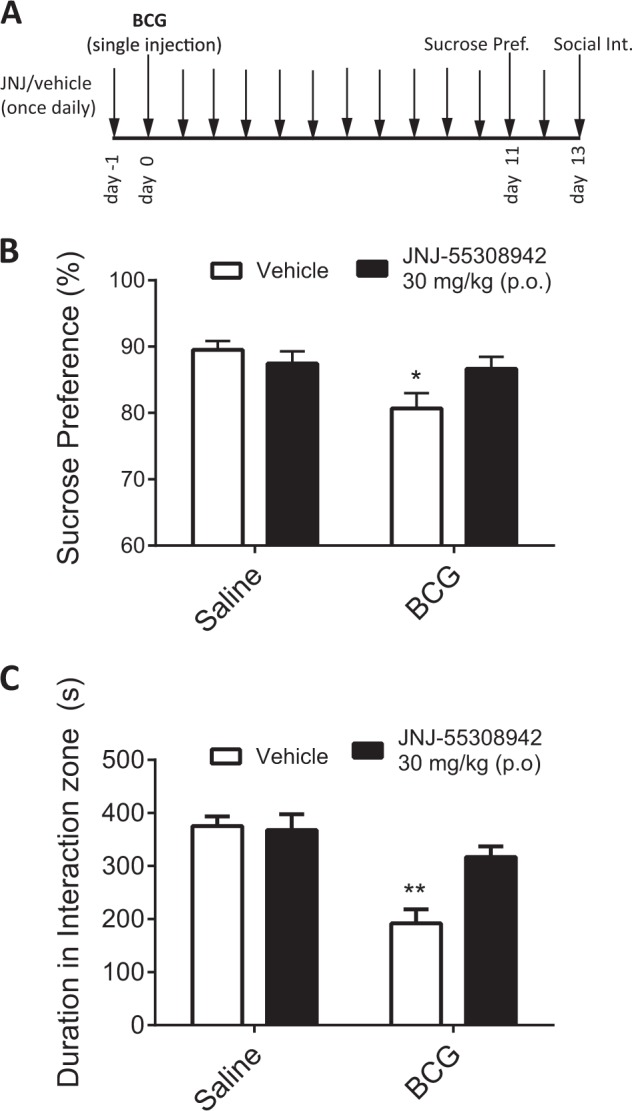
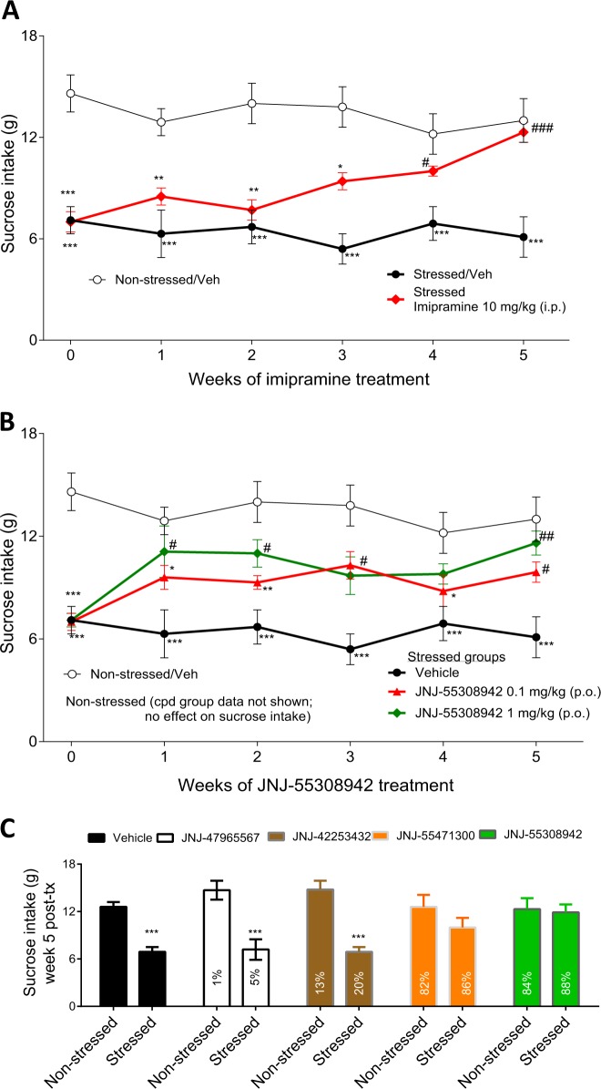
Emerging data continues to point towards a relationship between neuroinflammation and neuropsychiatric disorders. ATP-induced activation of P2X7 results in IL-1β release causing neuroinflammation and microglial activation. This study describes the in-vitro and in-vivo neuropharmacology of a novel brain-penetrant P2X7 antagonist, JNJ-55308942, currently in clinical development. JNJ-55308942 is a high-affinity, selective, brain-penetrant (brain/plasma of 1) P2X7 functional antagonist. In human blood and in mouse blood and microglia, JNJ-55308942 attenuated IL-1β release in a potent and concentration-dependent manner. After oral dosing, the compound exhibited both dose and concentration-dependent occupancy of rat brain P2X7 with an ED50 of 0.07 mg/kg. The P2X7 antagonist (3 mg/kg, oral) blocked Bz-ATP-induced brain IL-1β release in conscious rats, demonstrating functional effects of target engagement in the brain. JNJ-55308942 (30 mg/kg, oral) attenuated LPS-induced microglial activation in mice, assessed at day 2 after a single systemic LPS injection (0.8 mg/kg, i.p.), suggesting a role for P2X7 in microglial activation. In a model of BCG-induced depression, JNJ-55308942 dosed orally (30 mg/kg), reversed the BCG-induced deficits of sucrose preference and social interaction, indicating for the first time a role of P2X7 in the BCG model of depression, probably due to the neuroinflammatory component induced by BCG inoculation. Finally, in a rat model of chronic stress induced sucrose intake deficit, JNJ-55308942 reversed the deficit with concurrent high P2X7 brain occupancy as measured by autoradiography. This body of data demonstrates that JNJ-55308942 is a potent P2X7 antagonist, engages the target in brain, modulates IL-1β release and microglial activation leading to efficacy in two models of anhedonia in rodents.


Conflict of interest statement

Except Drs. Jason O’Connor and Mariusz Papp, all authors are employees of Johnson & Johnson. The authors declare no competing interests.

Figures

Fig. 1
Fig. 1
P2X7 antagonism attenuates IL-1β release and cell death of microglia. a Chemical structure of the P2X7 antagonist JNJ-55308942. b Secretion of IL-1β of microglia after pre-treatment with different concentrations of JNJ-55308942 in the presence of 100 ng/ml LPS followed by 380 μM Bz-ATP for additional 2 h and quantified by ELISA. c Cell number of microglia after pre-treatment with different concentrations of JNJ-55308942 in the presence of 380 μM Bz-ATP quantified by CCK-8 kit. The proportion of microglial cell death was obtained by normalizing to the condition of Bz-ATP treatment only. Data shown is mean ± standard deviation. Experiments were done in six replicates and repeated twice independently
Fig. 2
Fig. 2
Demonstration of target engagement of JNJ-55308942 in the brain and blood. a Dose vs. brain occupancy curve at 2 h post a single oral administration of JNJ-55308942 in the rat. Plasma and brain exposure of the compound for each dose is shown as an inset table. b Time course of rat brain P2X7 occupancy after a single 10 mg/kg dose (oral) of JNJ-55308942. Plasma and brain was harvested at various time points (X-axis) to monitor changes in occupancy and exposure (inset). c Bz-ATP induced increases in brain IL-1β levels as measured by microdialysis in freely moving rats. The increase in IL-1β was observed during the time of Bz-ATP infusion, as indicated by the shaded bar/line. The compound was administered at a dose of 3 mg/kg (oral), 4 h prior to Bz-ATP infusion. d Effect of a single 30 mg/kg (oral) dose of JNJ-55308942 on ex-vivo IL-1β release in mouse blood. Mice were sacrificed at various time points to assess exposure in the brain/plasma and to study the effect of the compound in the blood on LPS primed Bz-ATP stimulated IL-1β release (black closed circle). At least three animals were used to generate data for each symbol in the figure. p < 0.01 at the 5 h time point, Wilcoxon test
Fig. 3
Fig. 3
Systemic P2X7 antagonism alleviates LPS-induced microglial activation. a Mice (N = 4/group) received the compound (30 mg/kg, oral) 1 h before as well as 7 and 24 h after an i.p. injection of LPS (0.8 mg/kg, i.p.) or vehicle and were sacrificed 48 h after LPS injection. Brains were collected, dissociated and stained with conjugated antibodies for FACS. b Gating strategy for (c): after a series of gates to exclude events with inconsistent flow rate, debris, doublets, and cell clumps (not shown), remaining events were gated for live cells by their relative size (forward scatter, FSC) and negative Zombie dye signal. Microglia were identified as CD45 int, CD11b+, CD206- cells within the live cell gate, and the median of their relative signal for CD11b and other markers (not shown) was assessed. c Relative fluorescent signal of microglia cells for FSC (correlating with cell size), side-scatter (SSC, correlating with cell complexity), CD45-BV605, and CD11b-PE-Cy7. LPS treatment causes significant increases in all four parameters. JNJ-55308942 (JNJ) significantly attenuates the effect of LPS on FSC, CD45 surface expression and CD11b surface expression, whereas in vehicle-treated controls the compound shows no effect. d A second aliquot of the dissociated brains was stained with the intranuclear proliferation marker Ki-67. After a series of gates to exclude debris, doublets etc. (not shown) the population encompassing microglia and other brain-resident macrophages was identified by their intermediate CD45 signal and positive CD11b staining. The percentage of cells positive for Ki-67-BV421 was evaluated in (e) e Injection of LPS dramatically increases the percentage of proliferating (Ki-67-positive) cells within brain-resident macrophages. This effect is significantly alleviated by systemic P2X7 blockade (JNJ). Results are shown as % of the non-treated control group. Boxes show mean with upper and lower quartiles with whiskers depicting the total range of the data. ##p < 0.01, ###p < 0.001 LPS vs. VEH, * p < 0.05 CPD vs. CTRL (two-way ANOVA followed by Tukey-corrected multiple comparisons)
Fig. 4
Fig. 4
JNJ-55308942 is efficacious in a BCG-model of depression. a The experimental schematic is shown; JNJ-55308942 (JNJ, 30 mg/kg, oral) was dosed a day prior to a single administration of BCG and every day thereafter. Behavior was measured on days indicated. b Sucrose preference was measured as an index of anhedonia-like behavior. In vehicle-treated control mice, BCG precipitated a significant 10% reduction in sucrose preference during a 48 h probe at day 11 post challenge (p < 0.05). Two-factor ANOVA revealed a significant drug x BCG interaction, and post hoc analysis confirmed that drug-treated mice did not exhibit BCG-induced reduction in sucrose preference. Data represent mean ± SEM, n = 8–11 mice/group. c BCG disrupts social interaction in a P2X7 receptor dependent manner. BCG reduced the amount of time that vehicle-treated mice spent in the interaction zone proximal to a novel stranger mouse (p < 0.05). Drug treatment significantly attenuated the reduction in social investigation precipitated by BCG challenge (drug × BCG interaction, p < 0.02) Data represent mean + /- SEM, n = 8–11 mice/group
Fig. 5
Fig. 5
JNJ-55308942 is efficacious in a chronic stress model of depression. Chronic stress resulted in a deficit of sucrose intake as demonstrated by open (non-stressed) and closed (stressed) circles in all panels a, b. The deficit of sucrose intake in the stressed rats was maintained during the 5 weeks of vehicle treatment. The effect of imipramine (10 mg/kg, i.p.) is shown in panel a. Efficacy of JNJ-55308942 (0.1 and 1 mg/kg, oral) is shown in panel b. Drug treatment had no effect on non-stressed rats (data not shown). c Relationship between sucrose intake (Y axis), P2X7 antagonists dosed at 1 mg/kg (oral) for 5 weeks (JNJ-47965567: white bar; JNJ-42253432: brown bar; JNJ-55471300: orange bar; JNJ-55308942: green bar) and corresponding mean brain P2X7 occupancy shown as numbers (±SEM) within the bar. Occupancy (as shown by the numbers within the bar graphs) represents binding of the compound at 3 h post dose. Sucrose intake was measured weekly from N = 8 rats, divided across various treatment groups. Individual analysis of each doses revealed that in stressed animals the two doses of JNJ-55308942 caused significant treatment effect [0.1 mg: F(1,84) = 28.749; p < 0.001, 1 mg: F(1,84) = 38.766; p < 0.001]. When compared to the week zero scores, the increase of intakes caused by 0.1 mg/kg was apparent after already first week of treatment, but it reached significance at two time points (i.e., week 3 & 5) only. The dose of 1 mg/kg increased intakes also already after first week of treatment (p = 0.014) and, except for week 3 & 4, this effect was further enhanced in the following tests. *p < 0.05, **p < 0.01, ***p < 0.001; relative to vehicle-treated or drug-treated control groups. #p < 0.05, ## p < 0.01; relative to drug-treated stressed animals at week 0

References

    1. Najjar S, Pearlman DM, Alper K, Najjar A, Devinsky O. Neuroinflammation and psychiatric illness. J Neuroinflamm. 2013;10:43. - PMC - PubMed
    1. Reus GZ, Fries GR, Stertz L, Badawy M, Passos IC, Barichello T, et al. The role of inflammation and microglial activation in the pathophysiology of psychiatric disorders. Neuroscience. 2015;300:141–54. doi: 10.1016/j.neuroscience.2015.05.018. - DOI - PubMed
    1. He Y, Taylor N, Fourgeaud L, Bhattacharya A. The role of microglial P2X7: modulation of cell death and cytokine release. J Neuroinflamm. 2017;14:135. doi: 10.1186/s12974-017-0904-8. - DOI - PMC - PubMed
    1. Lord B, Ameriks MK, Wang Q, Fourgeaud L, Vliegen M, Verluyten W, et al. A novel radioligand for the ATP-gated ion channel P2X7: [3H] JNJ-54232334. Eur J Pharmacol. 2015;765:551–9. doi: 10.1016/j.ejphar.2015.09.026. - DOI - PubMed
    1. Bhattacharya A, Biber K. The microglial ATP-gated ion channel P2X7 as a CNS drug target. Glia. 2016;64:1772–87. doi: 10.1002/glia.23001. - DOI - PubMed

Publication types

MeSH terms