

Endoplasmic reticulum stress signalling - from basic mechanisms to clinical applications
- PMID: 30027602
- PMCID: PMC7379631
- DOI: 10.1111/febs.14608
Endoplasmic reticulum stress signalling - from basic mechanisms to clinical applications
Abstract
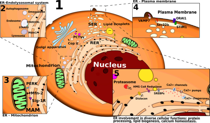
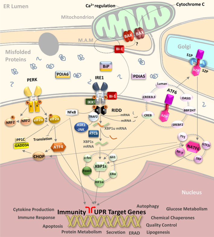
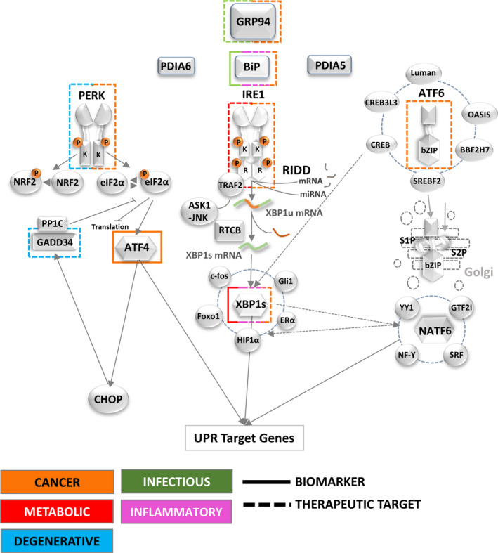
The endoplasmic reticulum (ER) is a membranous intracellular organelle and the first compartment of the secretory pathway. As such, the ER contributes to the production and folding of approximately one-third of cellular proteins, and is thus inextricably linked to the maintenance of cellular homeostasis and the fine balance between health and disease. Specific ER stress signalling pathways, collectively known as the unfolded protein response (UPR), are required for maintaining ER homeostasis. The UPR is triggered when ER protein folding capacity is overwhelmed by cellular demand and the UPR initially aims to restore ER homeostasis and normal cellular functions. However, if this fails, then the UPR triggers cell death. In this review, we provide a UPR signalling-centric view of ER functions, from the ER's discovery to the latest advancements in the understanding of ER and UPR biology. Our review provides a synthesis of intracellular ER signalling revolving around proteostasis and the UPR, its impact on other organelles and cellular behaviour, its multifaceted and dynamic response to stress and its role in physiology, before finally exploring the potential exploitation of this knowledge to tackle unresolved biological questions and address unmet biomedical needs. Thus, we provide an integrated and global view of existing literature on ER signalling pathways and their use for therapeutic purposes.
Keywords: endoplasmic reticulum; proteostasis; signalling pathway; stress.
© 2018 The Authors. The FEBS Journal published by John Wiley & Sons Ltd on behalf of Federation of European Biochemical Societies.
Figures
References
-
- Alberts B, Johnson A, Lewis J, Raff M, Roberts K & Walter P (2002) Molecular Biology of the Cell, 4th edn. Garland Science, New York, NY.
-
- Shibata Y, Voeltz GK & Rapoport TA (2006) Rough sheets and smooth tubules. Cell 126, 435–439. - PubMed
Publication types
MeSH terms
Associated data
- Actions
- Actions
- Actions
- Actions
LinkOut - more resources
Full Text Sources
Other Literature Sources
Research Materials
