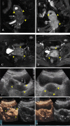
Contrast-enhanced ultrasonography vs MRI for evaluation of local invasion by cervical cancer
- PMID: 30028181
- PMCID: PMC6475950
- DOI: 10.1259/bjr.20170858
Contrast-enhanced ultrasonography vs MRI for evaluation of local invasion by cervical cancer
Abstract
Objective:: The purpose of this study is to compare contrast-enhanced ultrasound (CEUS) to MRI for evaluating local invasion of cervical cancer.
Methods:: A total of 108 patients with cervical cancer were included in this study. All the enrolled patients were Stage IIA2-IVB according to the International Federation of Obstetrics and Gynecology and treated with volumetric modulated arc therapy. Tumour size in different dimensions was compared between MRI and CEUS. The correlation coefficients (r) between MRI and CEUS for diagnosing local invasion, parametrial extension, and invasion to vagina, uterine corpus and adjacent organs were assessed.
Results:: Measurements by MRI and CEUS were strongly correlated in the three dimensions: left-right r = 0.84, craniocaudal r = 0.86 and anteroposterior r = 0.88. Vaginal and parametrial invasion were detected by both MRI and CEUS with moderate concordance, and invasion of uterine corpus, bladder and rectum with good concordance.
Conclusion:: CEUS is comparable to MRI for measuring tumour size, with good concordance for evaluating invasion of cervical cancer.
Advances in knowledge:: CEUS is a less expensive non-invasive modality for assessment of tumour size and invasion of cervical cancer.
Figures


Similar articles
-
Comparison of contrast-enhanced ultrasonography and magnetic resonance imaging in the evaluation of tumor size and local invasion of surgically treated cervical cancer.Abdom Radiol (NY). 2022 Aug;47(8):2928-2936. doi: 10.1007/s00261-022-03558-6. Epub 2022 Jun 7. Abdom Radiol (NY). 2022. PMID: 35670876
-
Interobserver agreement of transvaginal ultrasound and magnetic resonance imaging in local staging of cervical cancer.Ultrasound Obstet Gynecol. 2021 Nov;58(5):773-779. doi: 10.1002/uog.23662. Ultrasound Obstet Gynecol. 2021. PMID: 33915001 Free PMC article.
-
Agreement of two-dimensional and three-dimensional transvaginal ultrasound with magnetic resonance imaging in assessment of parametrial infiltration in cervical cancer.Ultrasound Obstet Gynecol. 2015 Apr;45(4):459-69. doi: 10.1002/uog.14637. Epub 2015 Mar 1. Ultrasound Obstet Gynecol. 2015. PMID: 25091827
-
Cervical carcinoma: computed tomography and magnetic resonance imaging for preoperative staging.Obstet Gynecol. 1995 Jul;86(1):43-50. doi: 10.1016/0029-7844(95)00109-5. Obstet Gynecol. 1995. PMID: 7784021 Review.
-
CT evaluation of cervical cancer: spectrum of disease.Radiographics. 2001 Sep-Oct;21(5):1155-68. doi: 10.1148/radiographics.21.5.g01se311155. Radiographics. 2001. PMID: 11553823 Review.
Cited by
-
Biplane transrectal ultrasonography plus ultrasonic elastosonography and contrast-enhanced ultrasonography in T staging of rectal cancer.BMC Cancer. 2020 Sep 7;20(1):862. doi: 10.1186/s12885-020-07369-0. BMC Cancer. 2020. PMID: 32894078 Free PMC article.
-
Comparing qSMI and qCEUS for assessing vascularization in uterine cervical cancer: operable versus non-operable group.Front Oncol. 2024 Aug 12;14:1380725. doi: 10.3389/fonc.2024.1380725. eCollection 2024. Front Oncol. 2024. PMID: 39188687 Free PMC article.
-
The safety of combining Endostar with concurrent chemoradiotherapy for the treatment of locally advanced cervical cancer and the evaluation of its anti-angiogenic effects via transrectal contrast-enhanced ultrasound.Front Oncol. 2025 May 8;15:1514425. doi: 10.3389/fonc.2025.1514425. eCollection 2025. Front Oncol. 2025. PMID: 40406255 Free PMC article.
-
Prognostic Value of Quantitative Perfusion Parameters by Enhanced Ultrasound in Endometrial Cancer.Med Sci Monit. 2019 Jan 10;25:298-304. doi: 10.12659/MSM.912782. Med Sci Monit. 2019. PMID: 30626861 Free PMC article.
-
The value of contrast-enhanced ultrasound in cervical cancer assessed in comparison with magnetic resonance imaging.Med Pharm Rep. 2024 Jul;97(3):255-262. doi: 10.15386/mpr-2746. Epub 2024 Jul 30. Med Pharm Rep. 2024. PMID: 39234448 Free PMC article.
References
-
- Amendola MA, Hricak H, Mitchell DG, Snyder B, Chi DS, Long HJ, et al. . Utilization of diagnostic studies in the pretreatment evaluation of invasive cervical cancer in the United States: results of intergroup protocol ACRIN 6651/GOG 183. J Clin Oncol 2005; 23: 7454–9. doi: 10.1200/JCO.2004.00.5397 - DOI - PubMed
Publication types
MeSH terms
Substances
LinkOut - more resources
Full Text Sources
Other Literature Sources
Medical

