. 2018 Nov 1;315(5):H1101-H1111.
doi: 10.1152/ajpheart.00209.2018. Epub 2018 Jul 20.

Caveolin-3 KO disrupts t-tubule structure and decreases t-tubular ICa density in mouse ventricular myocytes

Affiliations

Caveolin-3 KO disrupts t-tubule structure and decreases t-tubular ICa density in mouse ventricular myocytes

Simon M Bryant et al. Am J Physiol Heart Circ Physiol. .

Abstract

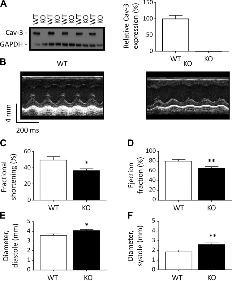
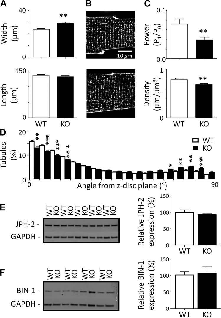
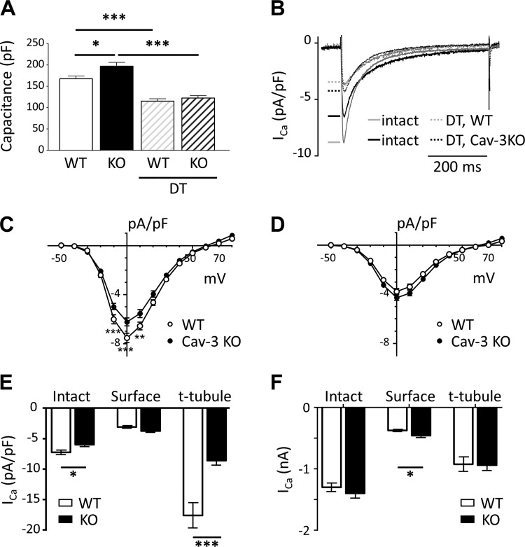
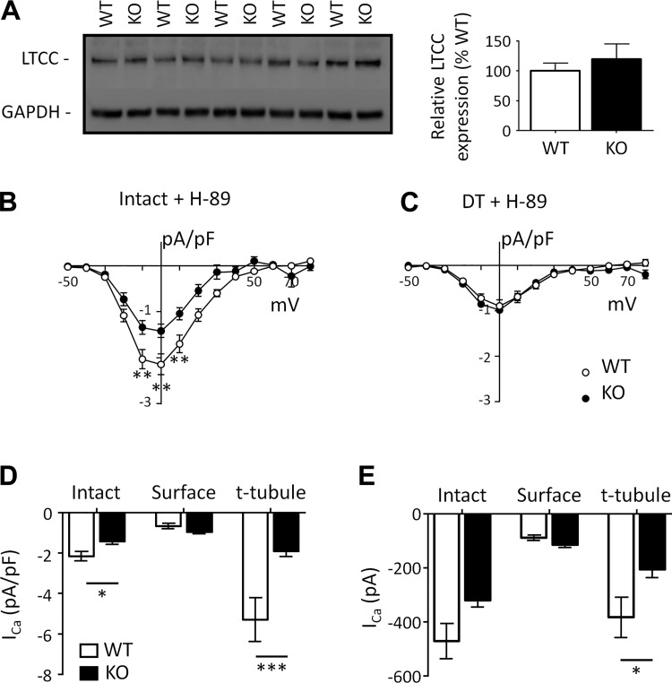
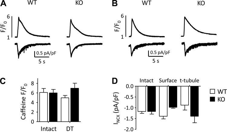
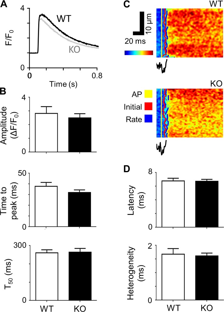
Caveolin-3 (Cav-3) is a protein that has been implicated in t-tubule formation and function in cardiac ventricular myocytes. In cardiac hypertrophy and failure, Cav-3 expression decreases, t-tubule structure is disrupted, and excitation-contraction coupling is impaired. However, the extent to which the decrease in Cav-3 expression underlies these changes is unclear. We therefore investigated the structure and function of myocytes isolated from the hearts of Cav-3 knockout (KO) mice. These mice showed cardiac dilatation and decreased ejection fraction in vivo compared with wild-type control mice. Isolated KO myocytes showed cellular hypertrophy, altered t-tubule structure, and decreased L-type Ca2+ channel current ( ICa) density. This decrease in density occurred predominantly in the t-tubules, with no change in total ICa, and was therefore a consequence of the increase in membrane area. Cav-3 KO had no effect on L-type Ca2+ channel expression, and C3SD peptide, which mimics the scaffolding domain of Cav-3, had no effect on ICa in KO myocytes. However, inhibition of PKA using H-89 decreased ICa at the surface and t-tubule membranes in both KO and wild-type myocytes. Cav-3 KO had no significant effect on Na+/Ca2+ exchanger current or Ca2+ release. These data suggest that Cav-3 KO causes cellular hypertrophy, thereby decreasing t-tubular ICa density. NEW & NOTEWORTHY Caveolin-3 (Cav-3) is a protein that inhibits hypertrophic pathways, has been implicated in the formation and function of cardiac t-tubules, and shows decreased expression in heart failure. This study demonstrates that Cav-3 knockout mice show cardiac dysfunction in vivo, while isolated ventricular myocytes show cellular hypertrophy, changes in t-tubule structure, and decreased t-tubular L-type Ca2+ current density, suggesting that decreased Cav-3 expression contributes to these changes in cardiac hypertrophy and failure.

Keywords: calcium current; calcium release; calcium transient; caveolin-3.


Conflict of interest statement

No conflicts of interest, financial or otherwise, are declared by the authors.

Figures

Fig. 1.
Fig. 1.
Caveolin-3 (Cav-3) and in vivo cardiac function. A: exemplar Western blots of Cav-3 (18 kDa) and GAPDH (37 kDa) in homogenates from wild-type (WT) and Cav-3 knockout (KO) mice (left) and mean densitometry data for Cav-3 Western blots (N = 5 animals in each group in duplicate; right). B: exemplar echocardiogram recordings from WT (left) and Cav-3 KO (right) mice. C–F: in vivo measurements of left ventricular cardiac function by echocardiography (WT: N = 6 and Cav-3 KO: N = 7). C: fractional shortening (in %). D: ejection fraction (in %). E: left ventricular internal diameter at diastole (in mm). F: left ventricular internal diameter at systole (in mm). *P < 0.05; **P < 0.01.
Fig. 2.
Fig. 2.
Morphology of isolated myocytes. A: cell width (top) and length (bottom) measured from bright-field images of myocytes from wild-type (WT) and caveolin-3 (Cav-3) knockout (KO) mice [WT n cells/N hearts (n/N): 100/4 and Cav-3 KO n/N: 30/4]. B: confocal images of t-tubules and surface sarcolemma stained with di-8-ANEPPs from representative WT (top) and Cav-3 KO (bottom) myocytes. C: t-tubule power (top) and density (bottom; WT n/N: 20/4 and Cav-3 KO n/N: 20/4). D: t-tubule orientation from the z-disk plane. E and F: Western blots of junctophilin-2 (JPH-2; 95 kDa; E) and bridging integrator-1 (BIN-1; 51 kDa; F) and GAPDH (37 kDa) (left) with corresponding mean data (N = 5 animals in each group in duplicate; right). *P < 0.05; **P < 0.01; ***P < 0.001.
Fig. 3.
Fig. 3.
Membrane capacitance and Ca2+ current (ICa). A: cell capacitance (in pF) of intact and detubulated (DT) myocytes used for electrophysiology from wild-type (WT) mice [intact n cells/N hearts (n/N): 35/13 and DT n/N = 38/13] and caveolin-3 (Cav-3) knockout (KO) mice (intact n/N: 39/17 and DT n/N: 43/14). B: exemplar records of ICa recorded at 0 mV from intact and DT WT and Cav-3 KO myocytes. C and D: mean ICa density-voltage relations from intact (C; WT n/N = 24/7 and Cav-3 KO n/N = 26/11) and DT (D; WT n/N = 28/9 and Cav-3 KO n/N = 31/9) WT and Cav-3 KO myocytes. E and F: mean ICa density (E) and absolute ICa (F) at 0 mV in intact and DT (“surface”) cells and calculated at the t-tubule membrane (“t-tubule”) for WT (open columns) and Cav-3 KO (solid columns) myocytes. *P < 0.05; **P < 0.01; ***P < 0.001.
Fig. 4.
Fig. 4.
Ca2+ current (ICa) in the presence of H-89. A: Western blots of the L-type Ca2+ channel (LTCC; 160 kDa) and GAPDH (37 kDa; left) and mean densitometry data (N = 5 animals in each group in duplicate; right). B and C: ICa density-voltage relations recorded in the presence of H-89 from intact [B; wild type (WT) n cells/N hearts (n/N): 16/9 and caveolin-3 (Cav-3) knockout (KO) n/N: 8/5] and detubulated (DT; C; WT n/N: 14/7 and Cav-3 KO n/N: 11/3) WT and Cav-3 KO myocytes. D and E: mean ICa density (D) and absolute ICa (E) at 0 mV in the presence of H-89 in intact and DT (“surface”) cells and calculated at the t-tubule membrane (“t-tubule”) for WT (open columns) and Cav-3 KO (solid columns) myocytes. *P < 0.05; **P < 0.01; ***P < 0.001.
Fig. 5.
Fig. 5.
Intracellular Ca2+ and Na+/Ca2+ exchanger current (INCX) during application of caffeine. A and B: exemplar records of the rise of intracellular Ca2+ (top) caused by application of 10 mM caffeine to intact (A) and detubulated (DT; B) wild-type (WT) and knockout (KO) myocytes and the accompanying inward currents (INCX; bottom). C: mean amplitude of the caffeine-induced rise of intracellular Ca2+ [intact: WT n cells/N hearts (n/N): 11/6 and Cav-3 KO n/N = 13/6; DT: WT n/N = 10/4, Cav-3 KO n/N = 12/3]. D: distribution of INCX density, calculated as previously described (26).
Fig. 6.
Fig. 6.
Systolic Ca2+ transients and local Ca2+ release. A: representative Ca2+ transients recorded from wild-type (WT) and caveolin-3 (Cav-3) knockout (KO) myocytes. B: Ca2+ transient amplitude, time to peak, and time to half decay (T50) measured from Ca2+ transients of WT [n cells/N hearts (n/N): 12/3] and Cav-3 KO (n/N = 14/3) cells. C: representative optical measurements of the rising phase of the Ca2+ transient and associated average t-tubular di-4-AN(F)EPPTEA signal. AP, action potential. D: mean latency and heterogeneity of sarcoplasmic reticulum Ca2+ release in WT (n/N = 10/3) and Cav-3 KO (n/N = 26/5) myocytes.

References

    1. Augustus AS, Buchanan J, Addya S, Rengo G, Pestell RG, Fortina P, Koch WJ, Bensadoun A, Abel ED, Lisanti MP. Substrate uptake and metabolism are preserved in hypertrophic caveolin-3 knockout hearts. Am J Physiol Heart Circ Physiol 295: H657–H666, 2008. doi:10.1152/ajpheart.00387.2008. - DOI - PMC - PubMed
    1. Balijepalli RC, Foell JD, Hall DD, Hell JW, Kamp TJ. Localization of cardiac L-type Ca2+ channels to a caveolar macromolecular signaling complex is required for β2-adrenergic regulation. Proc Natl Acad Sci USA 103: 7500–7505, 2006. doi:10.1073/pnas.0503465103. - DOI - PMC - PubMed
    1. Bers DM. Altered cardiac myocyte Ca regulation in heart failure. Physiology (Bethesda) 21: 380–387, 2006. doi:10.1152/physiol.00019.2006. - DOI - PubMed
    1. Boyett MR, Frampton JE, Kirby MS. The length, width and volume of isolated rat and ferret ventricular myocytes during twitch contractions and changes in osmotic strength. Exp Physiol 76: 259–270, 1991. doi:10.1113/expphysiol.1991.sp003492. - DOI - PubMed
    1. Brette F, Komukai K, Orchard CH. Validation of formamide as a detubulation agent in isolated rat cardiac cells. Am J Physiol Heart Circ Physiol 283: H1720–H1728, 2002. doi:10.1152/ajpheart.00347.2002. - DOI - PubMed

Publication types

MeSH terms

LinkOut - more resources