. 2018 Sep 25;12(9):8855-8866.
doi: 10.1021/acsnano.8b02805. Epub 2018 Jul 26.

Engineering a Rugged Nanoscaffold To Enhance Plug-and-Display Vaccination

Affiliations

Engineering a Rugged Nanoscaffold To Enhance Plug-and-Display Vaccination

Theodora U J Bruun et al. ACS Nano. .

Abstract

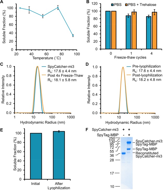
Nanoscale organization is crucial to stimulating an immune response. Using self-assembling proteins as multimerization platforms provides a safe and immunogenic system to vaccinate against otherwise weakly immunogenic antigens. Such multimerization platforms are generally based on icosahedral viruses and have led to vaccines given to millions of people. It is unclear whether synthetic protein nanoassemblies would show similar potency. Here we take the computationally designed porous dodecahedral i301 60-mer and rationally engineer this particle, giving a mutated i301 (mi3) with improved particle uniformity and stability. To simplify the conjugation of this nanoparticle, we employ a SpyCatcher fusion of mi3, such that an antigen of interest linked to the SpyTag peptide can spontaneously couple through isopeptide bond formation (Plug-and-Display). SpyCatcher-mi3 expressed solubly to high yields in Escherichia coli, giving more than 10-fold greater yield than a comparable phage-derived icosahedral nanoparticle, SpyCatcher-AP205. SpyCatcher-mi3 nanoparticles showed high stability to temperature, freeze-thaw, lyophilization, and storage over time. We demonstrate approximately 95% efficiency coupling to different transmission-blocking and blood-stage malaria antigens. Plasmodium falciparum CyRPA was conjugated to SpyCatcher-mi3 nanoparticles and elicited a high avidity antibody response, comparable to phage-derived virus-like particles despite their higher valency and RNA cargo. The simple production, precise derivatization, and exceptional ruggedness of this nanoscaffold should facilitate broad application for nanobiotechnology and vaccine development.

Keywords: bioconjugation; bionanotechnology; nanomedicine; protein engineering; self-assembly; vaccination; virus-like particle.


Conflict of interest statement

The authors declare the following competing financial interest(s): M.H. is an inventor on a patent regarding peptide targeting via spontaneous amide bond formation (EP2534484). M.H. and S.J.D. are SpyBiotech shareholders and consultants.

Figures

Figure 1
Figure 1
Design principle of rugged nanoscaffold platform. (a) Cartoon showing Plug-and-Display decoration of SpyCatcher-nanoparticles with a SpyTag-antigen and key issues for a robust platform. (b) Modification of the i301 scaffold. Two surface-exposed cysteines, C76 and C100 in i301 (yellow), were mutated to alanine to produce the mi3 nanoparticle platform. i301 is shown as the intact 60-mer and inset as a single monomer (based on the designed structure PyMOL file). The N-terminus is highlighted in blue. (c) mi3 increased the uniformity of the purified protein. Induction and purification gel, showing the additional disulfide-bonded species present for SpyCatcher-i301 but not SpyCatcher-mi3, analyzed by SDS-PAGE with Coomassie staining. (d) mi3 enhanced nanoscaffold yield. Purified protein yield (mean ±1 s.d., n = 3) was compared for SpyCatcher-mi3, SpyCatcher-i301, and SpyCatcher-AP205. (e) SpyCatcher-mi3 showed greater stability to aggregation. SpyCatcher-i301 or SpyCatcher-mi3 were stored for 5 weeks at 4 °C and analyzed by DLS.
Figure 2
Figure 2
Biophysical characterization of SpyCatcher-mi3 nanoparticles. (a) Size-exclusion chromatography of SpyCatcher-mi3 showed assembly into nanoparticles by absorbance at A280 (blue line), with comparison to molecular weight markers (gray line). (b) DLS determination of hydrodynamic radius (Rh) of SpyCatcher-mi3. (c) Negatively stained TEM image of SpyCatcher-mi3 nanoparticles. Scale bar 25 nm. (d) Size distribution of SpyCatcher-mi3 nanoparticles from TEM (n = 100).
Figure 3
Figure 3
SpyCatcher-mi3 nanoparticles efficiently reacted with a variety of antigens. Conjugation was performed with SpyCatcher-mi3 in a 1:3 ratio with antigen at 25 °C for 16 h, followed by analysis on SDS-PAGE with Coomassie staining. (a) SpyTag-MBP. (b) SpyTag-CIDR. (c) Pfs25-SpyTag. (d) CyRPA-SpyTag. (e) Coupling efficiency of SpyCatcher-mi3 with antigens, quantified by SDS-PAGE with Coomassie staining (mean of triplicate ±1 s.d.). (f) DLS characterization of SpyCatcher-mi3 with or without CyRPA-SpyTag conjugation (mean of triplicate ±1 s.d.).
Figure 4
Figure 4
SpyCatcher-mi3 nanoparticles were robust to heating, freezing, and lyophilization. (a) SpyCatcher-mi3 remained soluble after high temperatures. SpyCatcher-mi3 was incubated for 1 h at the indicated temperatures. Samples were centrifuged to remove aggregates, and soluble protein was quantified by densitometry (mean ±1 s.d., n = 3). (b) SpyCatcher-mi3 remained soluble after freeze–thaw. Soluble protein for SpyCatcher-mi3 was quantified, as in (a), after 1 or 4 cycles of freeze–thawing ±1 M trehalose (mean ±1 s.d., n = 3). (c) Freeze–thaw did not change nanoassembly. DLS of SpyCatcher-mi3 before and after four rounds of freeze–thaw without trehalose. (d) Lyophilization did not change nanoassembly. SpyCatcher-mi3 was analyzed by DLS before lyophilization or after lyophilization and reconstitution in the same volume. (e) SpyCatcher-mi3 soluble fraction, before and after lyophilization with reconstitution in the same buffer volume (mean ±1 s.d., n = 3). (f) SpyCatcher-mi3 retained reactivity after lyophilization. Reaction of SpyCatcher-mi3 with SpyTag-MBP at 25 °C for 16 h was analyzed by SDS-PAGE with Coomassie staining.
Figure 5
Figure 5
mi3 nanoassembly enhanced immunogenicity. (a) SpyCatcher-mi3 immunogen assembly. SpyCatcher-mi3 was conjugated with CyRPA-SpyTag, and unreacted antigen was removed by dialysis, as analyzed by SDS-PAGE with Coomassie staining. (b) SpyCatcher-AP205 immunogen assembly. SpyCatcher-AP205 was conjugated with CyRPA-SpyTag and analyzed as in (a). (c) Time course of immunization regimen. (d) Antibody titer to CyRPA after the prime. (e) Antibody titer to CyRPA after the boost. (f) Nanoassembly enhanced antibody avidity. The avidity of anti-CyRPA antibodies was assayed, based on resistance to NaSCN-induced dissociation. In each case, the value is plotted for each mouse. The horizontal bar represents the median and a Kruskal–Wallis test followed by Dunn’s multiple comparison post-test was performed. * p < 0.05; n.s. p > 0.05; n = 6.

References

    1. Kiessling L. L.; Gestwicki J. E.; Strong L. E. Synthetic Multivalent Ligands as Probes of Signal Transduction. Angew. Chem., Int. Ed. 2006, 45, 2348–2368. 10.1002/anie.200502794. - DOI - PMC - PubMed
    1. Mitragotri S.; Anderson D. G.; Chen X.; Chow E. K.; Ho D.; Kabanov A. V.; Karp J. M.; Kataoka K.; Mirkin C. A.; Petrosko S. H.; Shi J.; Stevens M. M.; Sun S.; Teoh S.; Venkatraman S. S.; Xia Y.; Wang S.; Gu Z.; Xu C. Accelerating the Translation of Nanomaterials in Biomedicine. ACS Nano 2015, 9, 6644–6654. 10.1021/acsnano.5b03569. - DOI - PMC - PubMed
    1. Zhang W.-B.; Sun F.; Tirrell D. A.; Arnold F. H. Controlling Macromolecular Topology with Genetically Encoded SpyTag-SpyCatcher Chemistry. J. Am. Chem. Soc. 2013, 135, 13988–13997. 10.1021/ja4076452. - DOI - PubMed
    1. Polka J. K.; Hays S. G.; Silver P. A. Building Spatial Synthetic Biology with Compartments, Scaffolds, and Communities. Cold Spring Harbor Perspect. Biol. 2016, 8, a024018.10.1101/cshperspect.a024018. - DOI - PMC - PubMed
    1. Uchida M.; McCoy K.; Fukuto M.; Yang L.; Yoshimura H.; Miettinen H. M.; LaFrance B.; Patterson D. P.; Schwarz B.; Karty J. A.; Prevelige P. E.; Lee B.; Douglas T. Modular Self-Assembly of Protein Cage Lattices for Multistep Catalysis. ACS Nano 2018, 12, 942–953. 10.1021/acsnano.7b06049. - DOI - PMC - PubMed

Publication types