Clinical Trial
. 2019 Jan;107(1):224-232.
doi: 10.1016/j.athoracsur.2018.06.015. Epub 2018 Jul 17.

A Clinical Trial of TumorGlow to Identify Residual Disease During Pleurectomy and Decortication

Affiliations
Clinical Trial

A Clinical Trial of TumorGlow to Identify Residual Disease During Pleurectomy and Decortication

Jarrod D Predina et al. Ann Thorac Surg. 2019 Jan.

Abstract

Background: Macroscopic complete resection can improve survival in a select group of patients with malignant pleural mesothelioma. During resection, differentiating residual tumor from inflammation or scar can be challenging. This trial evaluated near-infrared (NIR) intraoperative imaging using TumorGlow (a novel NIR imaging approach utilizing high-dose indocyanine green and delayed imaging) technology to improve detection of macroscopic residual disease.

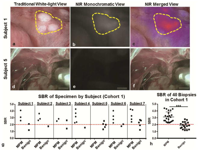
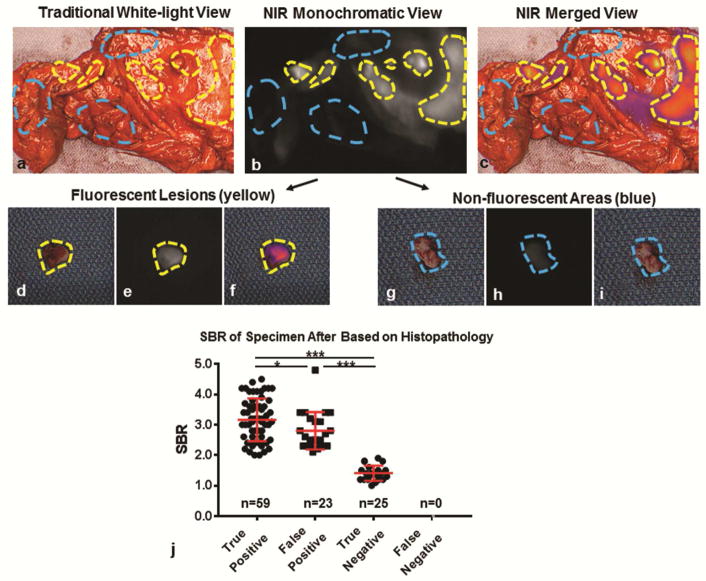
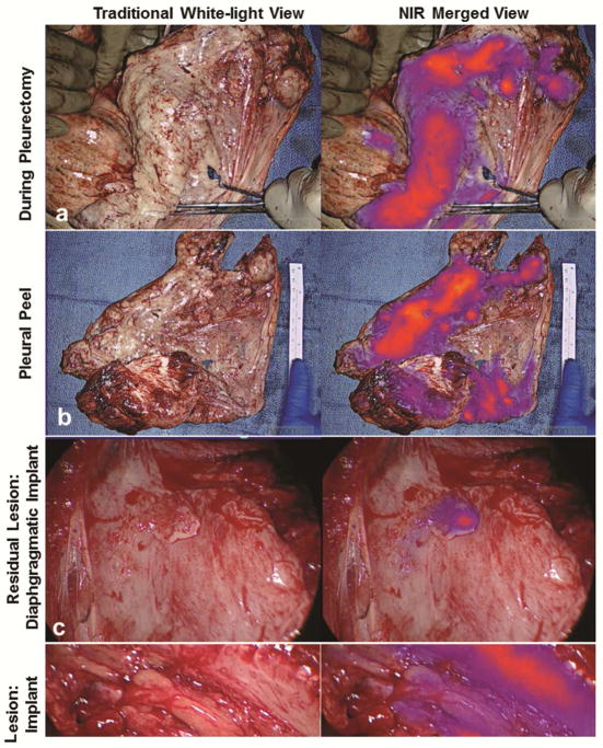
Methods: Twenty subjects were enrolled in an open-label clinical trial of NIR intraoperative imaging with TumorGlow (Indocyanine Green for Solid Tumors [NCT02280954]). Twenty-four hours before pleural biopsy or pleurectomy and decortication (P/D), patients received intravenous indocyanine green. All specimens identified during standard-of-care surgical resection and with NIR imaging underwent histopathologic profiling and correlative microscopic fluorescent tomographic evaluation. For subjects undergoing P/D (n = 13), the hemithorax was evaluated with NIR imaging during P/D to assess for residual disease. When possible, additional fluorescent lesions were resected.

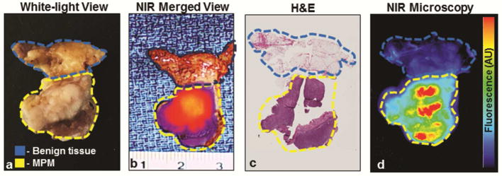
Results: Of 203 resected specimens submitted for evaluation, indocyanine green accumulated within 113 of 113 of resected mesothelioma specimens, with a mean signal-to-background fluorescence ratio of 3.1 (SD, 2.2 to 4.8). The mean signal-to-background fluorescence ratio of benign tissues was 2.2 (SD, 1.4 to 2.4), which was significantly lower than in malignant specimens (p = 0.001). NIR imaging identified occult macroscopic residual disease in 10 of 13 subjects. A median of 5.6 resectable residual deposits per patient (range, 0 to 11 deposits per patient), with a mean size of 0.3 cm (range, 0.1 to 1.5 cm), were identified.

Conclusions: TumorGlow for malignant pleural mesothelioma is safe and feasible. Excellent sensitivity allows for to reliable detection of macroscopic residual disease during cytoreductive surgical procedures.


Figures

Figure 1
Figure 1
FGS with ICG identified MPM during pleural biopsy: Subject 1: Representative example of subject in which high levels of fluorescence were observed during pleural biopsy. Subject 1, sample images of a cluster of 1mm MPM lesions (yellow gate) are displayed in a (a) traditional white-light view, (b) a monochromatic NIR view and (c) a merged NIR view. In Subject 5, there were no lesions identified during (d) white light thoracoscopy, nor during (d) NIR monochromatic or (e) merged NIR evaluation. (g) For each subject, the SBR of biopsied lesions were recorded and categorized based on final pathologic diagnosis. (h) The fluorescence (SBR) of biopsy proven MPM was significantly higher than benign lesions (p<0.0001).
Figure 2
Figure 2
ICG accumulates in resected MPM and displays fluorescence during cytoreductive surgery: Subject 12: Representative example of post-resection macroscopic and microscopic semi-quantitative fluorescence evaluation. Resected specimen were evaluated ex vivo using a combination of white-light views (a) and NIR views (b–c). Using NIR fluorescence patterns, fluorescent samples (yellow gate) and non-fluorescent samples (blue gate) were isolated, and submitted individually by a pathologist. As can be seen, fluorescent lesions were cleanly dissected from surrounding tissue (d–f), while non-fluorescent areas were also obtained (g–i). After obtaining a pathologic diagnosis for each submitted specimen (n=107), we plotted SBR for true-positives, false-positives, true-negatives and false-negatives. We found that the SBR of fluorescent MPM lesions (true positives) was higher than benign lesions which displayed fluorescence (false positives); p=0.03 (j). Both true positives and false positives displayed significantly higher fluorescence patterns than non-fluorescent benign tissues (true negatives); p<0.001. * - p<0.05; *** - p<0.0001
Figure 3
Figure 3
NIR Imaging with ICG accumulates preferentially in MPM. Specimens resected by standard-of-care approaches underwent macroscopic fluorescent profiling using a combination of standard white-light views (a), NIR views and NIR merged views (b). All resected specimens then underwent a series of histopathologic (c) and microscopic fluorescent analyses (d) to determine accuracy and patterns of dye accumulation. yellow gate—tumor by histopathologic review; blue gate—benign tissue by histopathologic analysis.
Figure 4
Figure 4
FGS with ICG identifies occult macroscopic residual disease following complete resection. Following macroscopic resection using white-light only, the ipsilateral hemithorax was then evaluated in NIR imaging to determine if residual disease was present. Data from Subject 10 is used to illustrate the workflow. The surgeon first completed P/D (a). All resected specimens underwent ex vivo macroscopic fluorescence evaluation (b). Next, the chest was reevaluated with NIR imaging to assess for residual disease. An example of a diaphragmatic implant (c) and an intercostal lesion (d) are provided. Both lesions were MPM by histopathologic review.

References

    1. Wolf AS, Flores RM. Current treatment of mesothelioma: Extrapleural pneumonectomy versus pleurectomy/decortication. Thorac Surg Clin. 2016;26(3):359–375. - PubMed
    1. Chan WH, Sugarbaker DJ, Burt BM. Intraoperative adjuncts for malignant pleural mesothelioma. Transl Lung Cancer Res. 2017;6(3):285–294. - PMC - PubMed
    1. Vogelzang NJ, Rusthoven JJ, Symanowski J, et al. Phase iii study of pemetrexed in combination with cisplatin versus cisplatin alone in patients with malignant pleural mesothelioma. J Clin Oncol. 2003;21(14):2636–2644. - PubMed
    1. Krug LM, Pass HI, Rusch VW, et al. Multicenter phase ii trial of neoadjuvant pemetrexed plus cisplatin followed by extrapleural pneumonectomy and radiation for malignant pleural mesothelioma. J Clin Oncol. 2009;27(18):3007–3013. - PMC - PubMed
    1. Baldini EH, Richards WG, Gill RR, et al. Updated patterns of failure after multimodality therapy for malignant pleural mesothelioma. J Thorac Cardiovasc Surg. 2015;149(5):1374–1381. - PMC - PubMed

Publication types

MeSH terms

Associated data