Review
. 2018 Jul 20;62(3):341-360.
doi: 10.1042/EBC20170104. Print 2018 Jul 20.

Mitochondrial dynamics: overview of molecular mechanisms

Affiliations
Review

Mitochondrial dynamics: overview of molecular mechanisms

Lisa Tilokani et al. Essays Biochem. .

Abstract

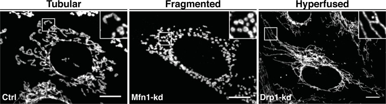
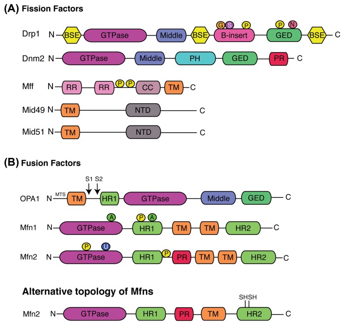
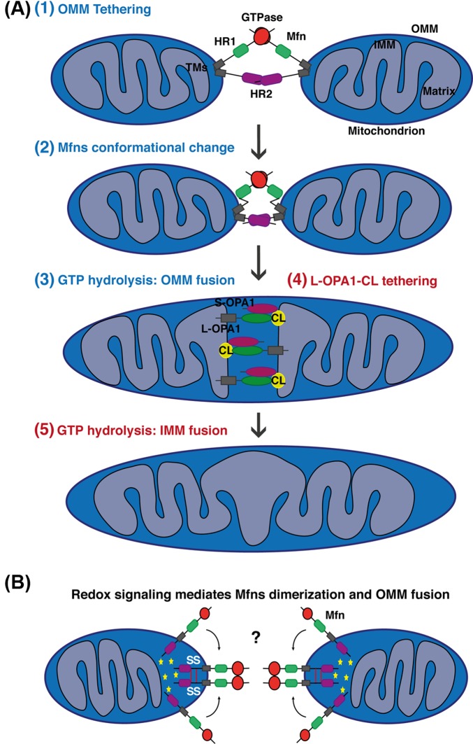
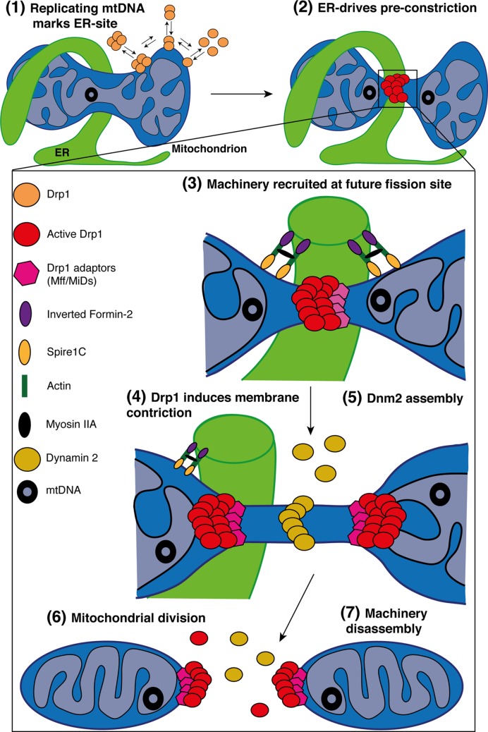
Mitochondria are highly dynamic organelles undergoing coordinated cycles of fission and fusion, referred as 'mitochondrial dynamics', in order to maintain their shape, distribution and size. Their transient and rapid morphological adaptations are crucial for many cellular processes such as cell cycle, immunity, apoptosis and mitochondrial quality control. Mutations in the core machinery components and defects in mitochondrial dynamics have been associated with numerous human diseases. These dynamic transitions are mainly ensured by large GTPases belonging to the Dynamin family. Mitochondrial fission is a multi-step process allowing the division of one mitochondrion in two daughter mitochondria. It is regulated by the recruitment of the GTPase Dynamin-related protein 1 (Drp1) by adaptors at actin- and endoplasmic reticulum-mediated mitochondrial constriction sites. Drp1 oligomerization followed by mitochondrial constriction leads to the recruitment of Dynamin 2 to terminate membrane scission. Inner mitochondrial membrane constriction has been proposed to be an independent process regulated by calcium influx. Mitochondrial fusion is driven by a two-step process with the outer mitochondrial membrane fusion mediated by mitofusins 1 and 2 followed by inner membrane fusion, mediated by optic atrophy 1. In addition to the role of membrane lipid composition, several members of the machinery can undergo post-translational modifications modulating these processes. Understanding the molecular mechanisms controlling mitochondrial dynamics is crucial to decipher how mitochondrial shape meets the function and to increase the knowledge on the molecular basis of diseases associated with morphology defects. This article will describe an overview of the molecular mechanisms that govern mitochondrial fission and fusion in mammals.

Keywords: Dynamin family; ER-Actin; Mitochondrial dynamics; Molecular Mechanisms; Regulation.


Conflict of interest statement

The authors declare that there are no competing interests associated with the manuscript.

Figures

Figure 1
Figure 1. The mitochondrial morphology network
Representative microscopy confocal images showing the different mitochondrial morphological aspects from control (Ctrl), Mfn1- and Drp1-Knockdown (Kd) mouse embryonic fibroblasts cells. Mitochondria are labelled with an anti-TOM20 antibody (OMM marker). Tubular, fragmented and hyperfused mitochondria are highlighted by zoomed areas (white squares); scale bars: 10 μm. Please note that the bright TOM20-positive structure in the zoom area of the Drp1-Kd is not a mitochondrial fragment but a mitochondria-derived vesicle [192].
Figure 2
Figure 2. Schematic representation of the structural elements of the fission and fusion proteins, and their associated post-translational modifications
Illustration of the core machinery proteins involved in (A) mitochondrial fission and (B) fusion. The classical model proposes that Mfns contain two transmembrane (TM) domains in between HR1 and HR2 domains. Alternatively, Mfns have been recently demonstrated to have only one TM that lies between the two HR domains. Cysteine residues, sensitive to oxidative stress are located in the C-terminal located in the IMS (only Mfn2 structural domains are represented but this new topology is also applicable to Mfn1). Domains are depicted in different colours. Identified location of post-translational modifications are indicated by P (Phosphorylation), N (S-nitrosylation), S (SUMOylation), G (O-GLcNAcylation), A (Acetylation) or U (Ubiquitination); BSE, bundle signalling elements; CC, coil-coil; GED, GTPase effector domain; HR, heptad repeat; MTS, mitochondrial targeting sequence; NTD, nucleotidyl transferase domains; PH, Pleckstrin homology; PR, Proline rich; RR, repeat regions; TM, transmembrane.
Figure 3
Figure 3. Simplified models for mitochondrial fusion in mammals
(A) Schematic representations of mitochondrial fusion, based on the Mfns topology suggesting two TM domains with both the HR1 and HR2 domains facing the cytosol. (1) The outer membrane of two opposing mitochondria are tethered by the interaction in trans of the HR2 and/or GTPase domains of Mfns. GTP binding or/and hydrolysis induce Mfns conformational change leading to mitochondrial docking and to an increase of membrane contact sites. For clarity reasons, not all of the recent suggested models leading to Mfns dimerization and conformational change are highlighted in the scheme. (3) Finally, GTPase-dependent power stroke or GTP-dependent oligomerization ensure OMM fusion. The composition of the OMM in phospholipids can also regulate this process. (4) Following OMM fusion, OPA1 and CL drive IMM fusion. The interaction between OPA1 and CL on either side of the membrane tethers the two IMM, which fuse following OPA1-depedent GTP hydrolysis (5). In this model, S-OPA1 has been shown to enhance OPA1–CL interaction and fusion. Please note that after OMM and IMM fusion, Mfn2 and OPA1, as membrane-bound proteins, are still present on the different membranes but are disassembled. (B) Schematic representations of OMM fusion based on the new metazoan Mfns topology suggesting only one TM placing the Mfn C-terminus in the IMS. Oxidized environment in the IMS (ROS production) and increase concentration of GSSG lead to the establishment of two disulphide bonds within the IMS domain. These redox-mediated disulphide modifications induce the dimerization and oligomerization of Mfns molecules which may promote tethering or GTPase activity required for OMM fusion. Interestingly, this redox-regulated Mfns oligomerization is a dynamic and reversible process. Yellow stars indicate an oxidized environment.
Figure 4
Figure 4. Simplified model for mitochondrial fission in mammals
Schematic representation of the multi-step processes required for mitochondria division. (1) In the matrix, replication of the mtDNA marks the site for ER-recruitment. In parallel, Drp1 oligomers are in constant balance between the cytosol and mitochondria. In addition, IMM constriction occurs at mitochondria–ER contacts in a Ca2+-dependent process, before Drp1 oligomerization and maturation. (2) Oligomeric forms of Drp1 accumulate at ER-sites where the pre-constriction of the membrane has been initiated. (3) The zoomed area highlights the factors regulating mitochondrial division. The ER-bound INF2 and mitochondrial Spire1C induce actin nucleation and polymerization at mitochondria–ER contact sites. The Myosin IIa may ensure actin cable contraction, providing the mechanical force to drive mitochondria pre-constriction. At these sites, MFF and MiDs recruit Drp1 where it oligomerizes in a ring-like structure and (4) GTP-hydrolysis leads to conformational change, enhancing pre-existing mitochondrial constriction. The composition of the OMM in phospholipids also regulates Drp1 assembly and activity. (5) Then, Dnm2 is recruited to Drp1-mediated mitochondrial constriction neck where it assembles and terminates membrane scission, (6) leading to two daughter mitochondria. (7) The mechanisms of disassembly of the fission machinery following division remain unclear but both adaptors and Drp1 are found at both mitochondrial tips after division.

References

    1. McBride H.M., Neuspiel M. and Wasiak S. (2006) Mitochondria: more than just a powerhouse. Curr. Biol. 16, R551–R560 10.1016/j.cub.2006.06.054 - DOI - PubMed
    1. Kamer K.J. and Mootha V.K. (2015) The molecular era of the mitochondrial calcium uniporter. Nat. Rev. Mol. Cell Biol. 16, 545–553 10.1038/nrm4039 - DOI - PubMed
    1. Nikoletopoulou V., Markaki M., Palikaras K. and Tavernarakis N. (2013) Crosstalk between apoptosis, necrosis and autophagy. Biochim. Biophys. Acta 1833, 3448–3459 10.1016/j.bbamcr.2013.06.001 - DOI - PubMed
    1. Rambold A.S. and Pearce E.L. (2018) Mitochondrial dynamics at the interface of immune cell metabolism and function. Trends Immunol. 39, 6–18 10.1016/j.it.2017.08.006 - DOI - PubMed
    1. Liesa M., Palacin M. and Zorzano A. (2009) Mitochondrial dynamics in mammalian health and disease. Physiol. Rev. 89, 799–845 10.1152/physrev.00030.2008 - DOI - PubMed

Publication types

MeSH terms

Substances